3AJ5

HA1 (HA33) subcomponent of botulinum type C progenitor toxin complexed with N-acetylgalactosamine, bound at site II


Experimental Data Snapshot

  • Method: X-RAY DIFFRACTION
  • Resolution: 1.80 Å
  • R-Value Free: 0.202 
  • R-Value Work: 0.165 
  • R-Value Observed: 0.167 

Starting Model: experimental
View more details

wwPDB Validation   3D Report Full Report


Ligand Structure Quality Assessment 


This is version 1.4 of the entry. See complete history


Literature

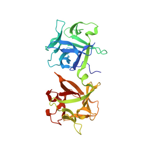
Molecular diversity of the two sugar-binding sites of the beta-trefoil lectin HA33/C (HA1) from Clostridium botulinum type C neurotoxin

Nakamura, T.Tonozuka, T.Ito, S.Takeda, Y.Sato, R.Matsuo, I.Ito, Y.Oguma, K.Nishikawa, A.

(2011) Arch Biochem Biophys 512: 69-77

  • DOI: https://doi.org/10.1016/j.abb.2011.05.012
  • Primary Citation of Related Structures:  
    3AJ5, 3AJ6

  • PubMed Abstract: 

    A critical role in internalizing the Clostridium botulinum neurotoxin into gastrointestinal cells is played by nontoxic components complexed with the toxin. One of the components, a β-trefoil lectin has been known as HA33 or HA1. The HA33 from C. botulinum type A (HA33/A) has been predicted to have a single sugar-binding site, while type C HA33 (HA33/C) has two sites. Here we constructed HA33/C mutants and evaluated the binding capacities of the individual sites through mucin-assay and isothermal titration calorimetry. The mutant W176A (site I knockout) had a K(d) value of 31.5mM for galactose (Gal) and 61.3mM for N-acetylgalactosamine (GalNAc), while the K(d) value for N-acetylneuraminic acid (Neu5Ac) was too high to be determined. In contrast, the double mutant N278A/Q279A (site II knockout) had a K(d) value of 11.8mM for Neu5Ac. We also determined the crystal structures of wild-type and the F179I mutant in complex with GalNAc at site II. The results suggest that site I of HA33/C is quite unique in that it mainly recognizes Neu5Ac, and site II seems less important for the lectin specificity. The architectures and the properties of the sugar-binding sites of HA33/C and HA33/A were shown to be drastically different.


  • Organizational Affiliation

    Department of Applied Biological Science, Tokyo University of Agriculture and Technology, 3-5-8, Saiwai-cho, Fuchu, Tokyo 183-8509, Japan.


Macromolecules
Find similar proteins by:  (by identity cutoff)  |  3D Structure
Entity ID: 1
MoleculeChains Sequence LengthOrganismDetailsImage
Main hemagglutinin component
A, B
286Clostridium botulinumMutation(s): 0 
UniProt
Find proteins for P0DPR0 (Clostridium botulinum C phage)
Explore P0DPR0 
Go to UniProtKB:  P0DPR0
Entity Groups  
Sequence Clusters30% Identity50% Identity70% Identity90% Identity95% Identity100% Identity
UniProt GroupP0DPR0
Sequence Annotations
Expand
  • Reference Sequence
Small Molecules
Ligands 1 Unique
IDChains Name / Formula / InChI Key2D Diagram3D Interactions
NGA
Query on NGA

Download Ideal Coordinates CCD File 
C [auth A],
D [auth B]
2-acetamido-2-deoxy-beta-D-galactopyranose
C8 H15 N O6
OVRNDRQMDRJTHS-JAJWTYFOSA-N
Experimental Data & Validation

Experimental Data

  • Method: X-RAY DIFFRACTION
  • Resolution: 1.80 Å
  • R-Value Free: 0.202 
  • R-Value Work: 0.165 
  • R-Value Observed: 0.167 
  • Space Group: C 1 2 1
Unit Cell:
Length ( Å )Angle ( ˚ )
a = 138.424α = 90
b = 62.187β = 104.63
c = 82.436γ = 90
Software Package:
Software NamePurpose
ADSCdata collection
MOLREPphasing
REFMACrefinement
HKL-2000data reduction
HKL-2000data scaling

Structure Validation

View Full Validation Report


Ligand Structure Quality Assessment 


Entry History 

Deposition Data

Revision History  (Full details and data files)

  • Version 1.0: 2011-06-01
    Type: Initial release
  • Version 1.1: 2011-07-13
    Changes: Version format compliance
  • Version 1.2: 2011-07-27
    Changes: Database references
  • Version 1.3: 2020-07-29
    Type: Remediation
    Reason: Carbohydrate remediation
    Changes: Data collection, Derived calculations, Structure summary
  • Version 1.4: 2023-11-01
    Changes: Data collection, Database references, Refinement description, Structure summary