


Crystal structure of Entamoeba histolytica methionine gamma-lyase 1
Karaki, T., Sato, D., Shimizu, A., Nozaki, T., Harada, S.To be published.
Experimental Data Snapshot
Starting Model: experimental
View more details
Entity ID: 1 | |||||
|---|---|---|---|---|---|
| Molecule | Chains | Sequence Length | Organism | Details | Image |
| Methionine gamma-lyase | 389 | Entamoeba histolytica | Mutation(s): 0 Gene Names: metg EC: 4.4.1.11 (PDB Primary Data), 2.5.1.160 (UniProt) | 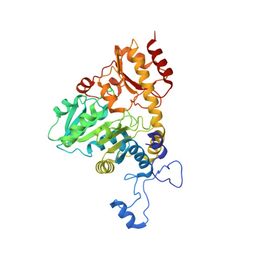 | |
UniProt | |||||
Entity Groups | |||||
| Sequence Clusters | 30% Identity50% Identity70% Identity90% Identity95% Identity100% Identity | ||||
| UniProt Group | Q86D28 | ||||
Sequence AnnotationsExpand | |||||
Reference Sequence | |||||
Entity ID: 2 | |||||
|---|---|---|---|---|---|
| Molecule | Chains | Sequence Length | Organism | Details | Image |
| Methionine gamma-lyase | 389 | Entamoeba histolytica | Mutation(s): 0 Gene Names: metg EC: 4.4.1.11 (PDB Primary Data), 2.5.1.160 (UniProt) | 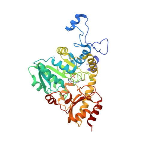 | |
UniProt | |||||
Entity Groups | |||||
| Sequence Clusters | 30% Identity50% Identity70% Identity90% Identity95% Identity100% Identity | ||||
| UniProt Group | Q86D28 | ||||
Sequence AnnotationsExpand | |||||
Reference Sequence | |||||
| Ligands 4 Unique | |||||
|---|---|---|---|---|---|
| ID | Chains | Name / Formula / InChI Key | 2D Diagram | 3D Interactions | |
| 2LM Download:Ideal Coordinates CCD File | E [auth A], H [auth B], M [auth D] | (2E)-2-[({3-hydroxy-2-methyl-5-[(phosphonooxy)methyl]pyridin-4-yl}methyl)imino]-4-(methylsulfanyl)butanoic acid C13 H19 N2 O7 P S RNHGWTJOZCEIDD-RVDMUPIBSA-N |  | ||
| MET Download:Ideal Coordinates CCD File | J [auth C] | METHIONINE C5 H11 N O2 S FFEARJCKVFRZRR-BYPYZUCNSA-N |  | ||
| SO4 Download:Ideal Coordinates CCD File | F [auth A], I [auth B], K [auth C], N [auth D] | SULFATE ION O4 S QAOWNCQODCNURD-UHFFFAOYSA-L |  | ||
| GOL Download:Ideal Coordinates CCD File | G [auth A], L [auth C] | GLYCEROL C3 H8 O3 PEDCQBHIVMGVHV-UHFFFAOYSA-N |  | ||
| Modified Residues 1 Unique | |||||
|---|---|---|---|---|---|
| ID | Chains | Type | Formula | 2D Diagram | Parent |
| LLP Query on LLP | C | L-PEPTIDE LINKING | C14 H22 N3 O7 P |  | LYS |
| Length ( Å ) | Angle ( ˚ ) |
|---|---|
| a = 99.086 | α = 90 |
| b = 85.332 | β = 101.9 |
| c = 114.331 | γ = 90 |
| Software Name | Purpose |
|---|---|
| DENZO | data reduction |
| SCALEPACK | data scaling |
| MOLREP | phasing |
| REFMAC | refinement |
| PDB_EXTRACT | data extraction |
| HKL-2000 | data collection |
| HKL-2000 | data reduction |
| HKL-2000 | data scaling |


