3A9L | pdb_00003a9l

Structure of Bacteriophage poly-gamma-glutamate hydrolase


Experimental Data Snapshot

  • Method: X-RAY DIFFRACTION
  • Resolution: 1.90 Å
  • R-Value Free: 
    0.315 (Depositor), 0.342 (DCC) 
  • R-Value Work: 
    0.281 (Depositor) 
  • R-Value Observed: 
    0.283 (Depositor) 

wwPDB Validation 3D Report Full Report

Validation slider image for 3A9L

This is version 1.3 of the entry. See complete history

Literature

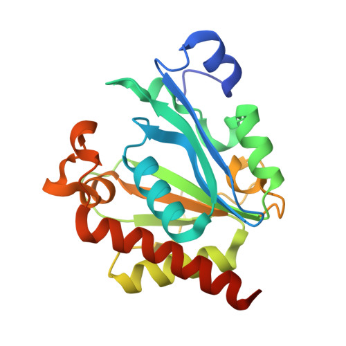
Crystal structure of bacteriophage PhiNIT1 zinc peptidase PghP that hydrolyzes gamma-glutamyl linkage of bacterial poly-gamma-glutamate

Fujimoto, Z.Kimura, K.

(2012) Proteins 80: 722-732

  • DOI: https://doi.org/10.1002/prot.23229
  • Primary Citation Related Structures: 
    3A9L

  • PubMed Abstract: 

    Poly-γ-glutamate hydrolase P (PghP) of Bacillus subtilis bacteriophage ΦNIT1 hydrolyzes the γ-glutamyl peptide linkage of extracellular poly-γ-glutamate produced by bacilli, which facilitates infection and propagation of phage progenies. Crystal structure of PghP was determined at a resolution of 1.9 Å. Structure of PghP was elucidated as a globular protein with an open α/β mixed core structure and a seven-stranded parallel/anti-parallel β-sheet. The β-sheet contained a core four-stranded parallel β-sheet. A zinc-binding motif, His-Glu-His, was identified at the C-terminal end of the β-sheet. Structure analysis demonstrated that PghP, which had not been previously classified into any peptidase/protease family due to lack of amino acid sequence similarity with known enzymes, had a catalytic center containing a zinc ion and an overall topology resembling mammalian carboxypeptidase A and related enzymes. Structural comparisons indicated important amino acid residues of PghP for catalysis and recognition of the γ-peptide bond of poly-γ-glutamate, which was confirmed by site-directed mutagenesis of PghP.


  • Organizational Affiliation
    • Biomolecular Research Unit, National Institute of Agrobiological Sciences, 2-1-2 Kannondai, Tsukuba, Ibaraki 305-8602, Japan. zui@affrc.go.jp

Macromolecule Content 

  • Total Structure Weight: 48.39 kDa 
  • Atom Count: 3,457 
  • Modeled Residue Count: 414 
  • Deposited Residue Count: 432 
  • Unique protein chains: 1

Macromolecules

Find similar proteins by:|  3D Structure
Entity ID: 1
MoleculeChains  Sequence LengthOrganismDetailsImage
Poly-gamma-glutamate hydrolase
A, B
216Bacillus phage phiNIT1Mutation(s): 0 
Gene Names: pghP
UniProt
Find proteins for Q852V1 (Bacillus phage phiNIT1)
Explore Q852V1 
Go to UniProtKB:  Q852V1
Entity Groups
Sequence Clusters30% Identity50% Identity70% Identity90% Identity95% Identity100% Identity
UniProt GroupQ852V1
Sequence Annotations
Expand
Reference Sequence

Experimental Data & Validation

Experimental Data

  • Method: X-RAY DIFFRACTION
  • Resolution: 1.90 Å
  • R-Value Free:  0.315 (Depositor), 0.342 (DCC) 
  • R-Value Work:  0.281 (Depositor) 
  • R-Value Observed: 0.283 (Depositor) 
Space Group: P 32
Unit Cell:
Length ( Å )Angle ( ˚ )
a = 86.809α = 90
b = 86.809β = 90
c = 85.116γ = 120
Software Package:
Software NamePurpose
UGUI@PFdata collection
SOLVEphasing
REFMACrefinement
HKL-2000data reduction
HKL-2000data scaling

Structure Validation

View Full Validation Report


Entry History 

Deposition Data

Revision History  (Full details and data files)

  • Version 1.0: 2010-11-10
    Type: Initial release
  • Version 1.1: 2011-07-13
    Changes: Version format compliance
  • Version 1.2: 2012-02-29
    Changes: Database references
  • Version 1.3: 2024-03-13
    Changes: Data collection, Database references, Derived calculations