3A7R | pdb_00003a7r

Crystal structure of E. coli lipoate-protein ligase A in complex with lipoyl-AMP.


Experimental Data Snapshot

  • Method: X-RAY DIFFRACTION
  • Resolution: 2.05 Å
  • R-Value Free: 
    0.204 (Depositor), 0.187 (DCC) 
  • R-Value Work: 
    0.176 (Depositor) 
  • R-Value Observed: 
    0.177 (Depositor) 

Starting Model: experimental
View more details

wwPDB Validation 3D Report Full Report

Validation slider image for 3A7R

Ligand Structure Quality Assessment 


This is version 1.2 of the entry. See complete history

Literature

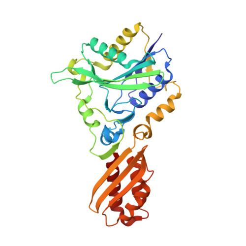
Global conformational change associated with the two-step reaction catalyzed by Escherichia coli lipoate-protein ligase A.

Fujiwara, K.Maita, N.Hosaka, H.Okamura-Ikeda, K.Nakagawa, A.Taniguchi, H.

(2010) J Biological Chem 285: 9971-9980

  • DOI: https://doi.org/10.1074/jbc.M109.078717
  • Primary Citation Related Structures: 
    3A7A, 3A7L, 3A7R, 3A7U

  • PubMed Abstract: 

    Lipoate-protein ligase A (LplA) catalyzes the attachment of lipoic acid to lipoate-dependent enzymes by a two-step reaction: first the lipoate adenylation reaction and, second, the lipoate transfer reaction. We previously determined the crystal structure of Escherichia coli LplA in its unliganded form and a binary complex with lipoic acid (Fujiwara, K., Toma, S., Okamura-Ikeda, K., Motokawa, Y., Nakagawa, A., and Taniguchi, H. (2005) J Biol. Chem. 280, 33645-33651). Here, we report two new LplA structures, LplA.lipoyl-5'-AMP and LplA.octyl-5'-AMP.apoH-protein complexes, which represent the post-lipoate adenylation intermediate state and the pre-lipoate transfer intermediate state, respectively. These structures demonstrate three large scale conformational changes upon completion of the lipoate adenylation reaction: movements of the adenylate-binding and lipoate-binding loops to maintain the lipoyl-5'-AMP reaction intermediate and rotation of the C-terminal domain by about 180 degrees . These changes are prerequisites for LplA to accommodate apoprotein for the second reaction. The Lys(133) residue plays essential roles in both lipoate adenylation and lipoate transfer reactions. Based on structural and kinetic data, we propose a reaction mechanism driven by conformational changes.


  • Organizational Affiliation
    • Institute for Enzyme Research, University of Tokushima, Kuramotocho 3-chome, Tokushima 770-8503. Electronic address: fujiwara@ier.tokushima-u.ac.jp.

Macromolecule Content 

  • Total Structure Weight: 38.78 kDa 
  • Atom Count: 3,224 
  • Modeled Residue Count: 337 
  • Deposited Residue Count: 337 
  • Unique protein chains: 1

Macromolecules

Find similar proteins by:|  3D Structure
Entity ID: 1
MoleculeChains  Sequence LengthOrganismDetailsImage
Lipoate-protein ligase A337Escherichia coli K-12Mutation(s): 0 
Gene Names: lplAyjjFb4386JW4349
EC: 6.3.4 (PDB Primary Data), 6.3.1.20 (UniProt)
UniProt
Find proteins for P32099 (Escherichia coli (strain K12))
Explore P32099 
Go to UniProtKB:  P32099
Entity Groups
Sequence Clusters30% Identity50% Identity70% Identity90% Identity95% Identity100% Identity
UniProt GroupP32099
Sequence Annotations
Expand
Reference Sequence

Small Molecules

Experimental Data & Validation

Experimental Data

  • Method: X-RAY DIFFRACTION
  • Resolution: 2.05 Å
  • R-Value Free:  0.204 (Depositor), 0.187 (DCC) 
  • R-Value Work:  0.176 (Depositor) 
  • R-Value Observed: 0.177 (Depositor) 
Space Group: P 61 2 2
Unit Cell:
Length ( Å )Angle ( ˚ )
a = 102.298α = 90
b = 102.298β = 90
c = 221.252γ = 120
Software Package:
Software NamePurpose
REFMACrefinement

Structure Validation

View Full Validation Report


Ligand Structure Quality Assessment 


Entry History 

Deposition Data

Revision History  (Full details and data files)

  • Version 1.0: 2010-01-19
    Type: Initial release
  • Version 1.1: 2011-07-13
    Changes: Version format compliance
  • Version 1.2: 2023-11-01
    Changes: Data collection, Database references, Derived calculations, Refinement description