3A7K | pdb_00003a7k

Crystal structure of halorhodopsin from Natronomonas pharaonis


Experimental Data Snapshot

  • Method: X-RAY DIFFRACTION
  • Resolution: 2.00 Å
  • R-Value Free: 
    0.274 (Depositor), 0.274 (DCC) 
  • R-Value Work: 
    0.249 (Depositor), 0.249 (DCC) 

Starting Model: experimental
View more details

wwPDB Validation 3D Report Full Report

Validation slider image for 3A7K

Ligand Structure Quality Assessment 


This is version 1.3 of the entry. See complete history

Literature

Crystal Structure of the Light-Driven Chloride Pump Halorhodopsin from Natronomonas pharaonis.

Kouyama, T.Kanada, S.Takeguchi, Y.Narusawa, A.Murakami, M.Ihara, K.

(2010) J Mol Biology 396: 564-579

  • DOI: https://doi.org/10.1016/j.jmb.2009.11.061
  • Primary Citation Related Structures: 
    3A7K

  • PubMed Abstract: 

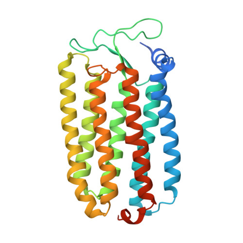
    The light-driven chloride pump halorhodopsin from Natronomonas pharaonis (phR) crystallised into the monoclinic space group C2, with a phR trimer per the asymmetric unit. Diffraction data at 2.0-A resolution showed that the carotenoid bacterioruberin binds to crevices between adjacent protein subunits in the trimeric assembly. Besides seven transmembrane helices (A to G) that characterise archaeal rhodopsins, the phR protomer possesses an amphipathic alpha-helix (A') at the N-terminus. This helix, together with a long loop between helices B and C, forms a hydrophobic cap that covers the extracellular surface and prevents a rapid ion exchange between the active centre and the extracellular medium. The retinal bound to Lys256 in helix G takes on an all-trans configuration with the Schiff base being hydrogen-bonded to a water molecule. The Schiff base also interacts with Asp252 and a chloride ion, the latter being fixed by two polar groups (Thr126 and Ser130) in helix C. In the anion uptake pathway, four ionisable residues (Arg123, Glu234, Arg176 and His100) and seven water molecules are aligned to form a long hydrogen-bonding network. Conversely, the cytoplasmic half is filled mostly by hydrophobic residues, forming a large energetic barrier against the transport of anion. The height of this barrier would be lowered substantially if the cytoplasmic half functions as a proton/HCl antiporter. Interestingly, there is a long cavity extending from the main-chain carbonyl of Lys256 to Thr71 in helix B. This cavity, which is commonly seen in halobacterial light-driven proton pumps, is one possible pathway that is utilised for a water-mediated proton transfer from the cytoplasmic medium to the anion, which is relocated to the cytoplasmic channel during the photocycle.


  • Organizational Affiliation
    • Department of Physics, Graduate School of Science, Nagoya University, Chikusa-ku, Nagoya 464-8602, Japan. kouyama@bio.phys.nagoya-u.ac.jp

Macromolecule Content 

  • Total Structure Weight: 110.3 kDa 
  • Atom Count: 6,678 
  • Modeled Residue Count: 776 
  • Deposited Residue Count: 873 
  • Unique protein chains: 1

Macromolecules

Find similar proteins by:|  3D Structure
Entity ID: 1
MoleculeChains  Sequence LengthOrganismDetailsImage
HalorhodopsinA,
B,
C [auth D]
291Natronomonas pharaonis DSM 2160Mutation(s): 0 

Small Molecules

Ligands 6 Unique
IDChains Name / Formula / InChI Key2D Diagram3D Interactions
L3P

Query on L3P


Download:Ideal Coordinates CCD File
BA [auth D]
I [auth A]
J [auth A]
K [auth A]
L [auth A]
BA [auth D],
I [auth A],
J [auth A],
K [auth A],
L [auth A],
M [auth A],
N [auth A],
O [auth A],
S [auth B]
2,3-DI-O-PHYTANLY-3-SN-GLYCERO-1-PHOSPHORYL-3'-SN-GLYCEROL-1'-PHOSPHATE
C46 H94 O11 P2
TZXJQSKPTCRGCA-VZSPAKCESA-L
22B

Query on 22B


Download:Ideal Coordinates CCD File
AA [auth D],
E [auth A],
R [auth B]
BACTERIORUBERIN
C50 H76 O4
UVCQMCCIAHQDAF-CUMPQFAQSA-N
L1P

Query on L1P


Download:Ideal Coordinates CCD File
F [auth A],
V [auth B],
X [auth B]
3-PHOSPHORYL-[1,2-DI-PHYTANYL]GLYCEROL
C43 H89 O6 P
UKQGAMWGTOTQPC-ALOLAALWSA-N
L2P

Query on L2P


Download:Ideal Coordinates CCD File
FA [auth D]
G [auth A]
GA [auth D]
H [auth A]
Y [auth B]
FA [auth D],
G [auth A],
GA [auth D],
H [auth A],
Y [auth B],
Z [auth B]
2,3-DI-PHYTANYL-GLYCEROL
C43 H88 O3
ISDBCJSGCHUHFI-UMZPFTBHSA-N
RET

Query on RET


Download:Ideal Coordinates CCD File
CA [auth D],
D [auth A],
T [auth B]
RETINAL
C20 H28 O
NCYCYZXNIZJOKI-OVSJKPMPSA-N
CL

Query on CL


Download:Ideal Coordinates CCD File
DA [auth D]
EA [auth D]
P [auth A]
Q [auth A]
U [auth B]
DA [auth D],
EA [auth D],
P [auth A],
Q [auth A],
U [auth B],
W [auth B]
CHLORIDE ION
Cl
VEXZGXHMUGYJMC-UHFFFAOYSA-M

Experimental Data & Validation

Experimental Data

  • Method: X-RAY DIFFRACTION
  • Resolution: 2.00 Å
  • R-Value Free:  0.274 (Depositor), 0.274 (DCC) 
  • R-Value Work:  0.249 (Depositor), 0.249 (DCC) 
Space Group: C 1 2 1
Unit Cell:
Length ( Å )Angle ( ˚ )
a = 151.83α = 90
b = 99.77β = 127.67
c = 99.27γ = 90
Software Package:
Software NamePurpose
ADSCdata collection
CNSrefinement
MOSFLMdata reduction
SCALAdata scaling
CNSphasing

Structure Validation

View Full Validation Report


Ligand Structure Quality Assessment 


Entry History 

Deposition Data

  • Released Date: 2009-12-15 
  • Deposition Author(s): Kouyama, T.

Revision History  (Full details and data files)

  • Version 1.0: 2009-12-15
    Type: Initial release
  • Version 1.1: 2011-07-13
    Changes: Version format compliance
  • Version 1.2: 2023-11-01
    Changes: Data collection, Database references, Derived calculations, Refinement description
  • Version 1.3: 2024-10-23
    Changes: Structure summary