3A77 | pdb_00003a77

The crystal structure of phosphorylated IRF-3


Experimental Data Snapshot

  • Method: X-RAY DIFFRACTION
  • Resolution: 1.80 Å
  • R-Value Free: 
    0.221 (Depositor), 0.220 (DCC) 
  • R-Value Work: 
    0.194 (Depositor), 0.194 (DCC) 
  • R-Value Observed: 
    0.197 (Depositor) 

Starting Model: experimental
View more details

wwPDB Validation 3D Report Full Report

Validation slider image for 3A77

This is version 1.3 of the entry. See complete history

Literature

Ser386 phosphorylation of transcription factor IRF-3 induces dimerization and association with CBP/p300 without overall conformational change.

Takahasi, K.Horiuchi, M.Fujii, K.Nakamura, S.Noda, N.N.Yoneyama, M.Fujita, T.Inagaki, F.

(2010) Genes Cells 15: 901-910

  • DOI: https://doi.org/10.1111/j.1365-2443.2010.01427.x
  • Primary Citation Related Structures: 
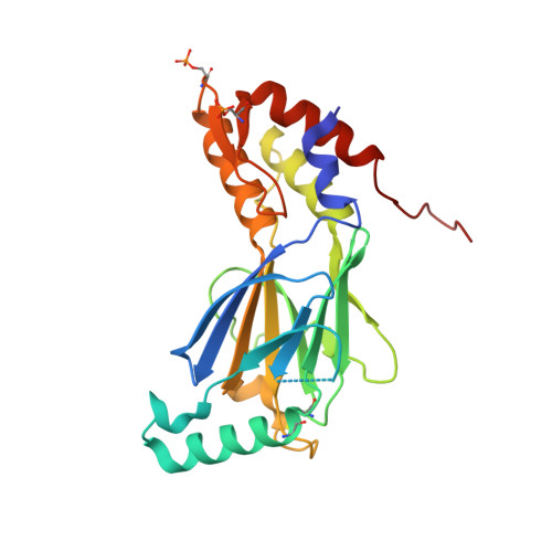
    3A77

  • PubMed Abstract: 

    The transcription factor IRF-3 is activated by microbial invasions and produces a variety of cytokines including type-I interferon. Upon microbial infection, IRF-3 is phosphorylated at its C-terminal regulatory domain, then oligomerized, translocated into the nucleus, and here it binds to CBP/p300. Although a number of studies have been reported investigating the activation mechanism of IRF-3, there are a number of unresolved issues, especially on the phosphorylation sites, the oligomerization process and the binding mechanism with CBP/p300. In this report, the phosphorylated IRF-3 regulatory domain (IRF-3 RD) was prepared using the kinase IKK-i, and the active form of phosphorylated IRF-3 RD was identified. The paper also reports the crystal structure of the active form of the phosphorylated IRF-3 RD. Furthermore, the phosphorylation of Ser386 was found to be essential for its dimerization and binding with CBP/p300 using mutational analysis and mass spectrometry. Thus, we conclude that the phosphorylation of Ser386 is essential for activation of IRF-3.


  • Organizational Affiliation
    • Department of Structural Biology, Graduate School of Pharmaceutical Sciences, Hokkaido University, N-12W-6 Kita-ku, Sapporo 060-0812, Japan.

Macromolecule Content 

  • Total Structure Weight: 108.42 kDa 
  • Atom Count: 8,328 
  • Modeled Residue Count: 929 
  • Deposited Residue Count: 968 
  • Unique protein chains: 1

Macromolecules

Find similar proteins by:|  3D Structure
Entity ID: 1
MoleculeChains  Sequence LengthOrganismDetailsImage
Interferon regulatory factor 3
A, B, C, D
242Homo sapiensMutation(s): 0 
Gene Names: IRF3
UniProt & NIH Common Fund Data Resources
Find proteins for Q14653 (Homo sapiens)
Explore Q14653 
Go to UniProtKB:  Q14653
PHAROS:  Q14653
GTEx:  ENSG00000126456 
Entity Groups
Sequence Clusters30% Identity50% Identity70% Identity90% Identity95% Identity100% Identity
UniProt GroupQ14653
Sequence Annotations
Expand
Reference Sequence

Experimental Data & Validation

Experimental Data

  • Method: X-RAY DIFFRACTION
  • Resolution: 1.80 Å
  • R-Value Free:  0.221 (Depositor), 0.220 (DCC) 
  • R-Value Work:  0.194 (Depositor), 0.194 (DCC) 
  • R-Value Observed: 0.197 (Depositor) 
Space Group: P 21 21 2
Unit Cell:
Length ( Å )Angle ( ˚ )
a = 163.469α = 90
b = 102.459β = 90
c = 68.921γ = 90
Software Package:
Software NamePurpose
HKL-2000data collection
CNSrefinement
HKL-2000data reduction
HKL-2000data scaling
CNSphasing

Structure Validation

View Full Validation Report


Entry History 

Deposition Data

Revision History  (Full details and data files)

  • Version 1.0: 2010-08-04
    Type: Initial release
  • Version 1.1: 2011-07-13
    Changes: Version format compliance
  • Version 1.2: 2023-11-01
    Changes: Data collection, Database references, Derived calculations, Refinement description
  • Version 1.3: 2024-10-30
    Changes: Structure summary