3A71 | pdb_00003a71

High resolution structure of Penicillium chrysogenum alpha-L-arabinanase


Experimental Data Snapshot

  • Method: X-RAY DIFFRACTION
  • Resolution: 1.14 Å
  • R-Value Free: 
    0.128 (Depositor), 0.105 (DCC) 
  • R-Value Work: 
    0.104 (DCC) 
  • R-Value Observed: 
    0.107 (Depositor) 

wwPDB Validation 3D Report Full Report

Validation slider image for 3A71

This is version 1.2 of the entry. See complete history

Literature

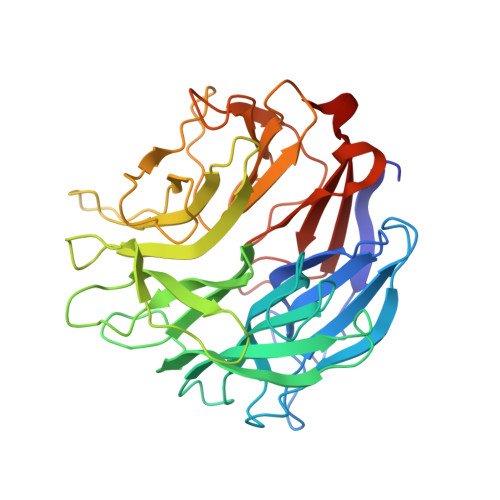
High-resolution structure of exo-arabinanase from Penicillium chrysogenum

Sogabe, Y.Kitatani, T.Yamaguchi, A.Kinoshita, T.Adachi, H.Takano, K.Inoue, T.Mori, Y.Matsumura, H.Sakamoto, T.Tada, T.

(2011) Acta Crystallogr D Biol Crystallogr 67: 415-422

  • DOI: https://doi.org/10.1107/S0907444911006299
  • Primary Citation Related Structures: 
    3A71, 3A72

  • PubMed Abstract: 

    Arabinanase Abnx from Penicillium chrysogenum 31B, which belongs to the GH93 family, releases arabinobiose from the nonreducing terminus of α-1,5-L-arabinan, which is distributed in the primary cell walls of higher plants. Crystal structures of Abnx and of its complex with arabinobiose were determined at the high resolutions of 1.14 Å to an R(work) of 10.7% (R(free) = 12.8%) and 1.04 Å to an R(work) of 10.4% (R(free) = 12.5%). Abnx has a six-bladed β-propeller fold with a typical ring-closure mode called `Velcro', in which the last four-stranded β-sheet is completed by the incorporation of a strand from the N-terminus. Catalytic residues which act as a nucleophile and an acid/base were proposed from the structures and confirmed by site-directed mutagenesis. The substrate-binding groove is enclosed at one end by two residues, Glu64 and Tyr66, which contribute to the recognition of the nonreducing chain end of the polysaccharide. A comparison with the related enzyme Arb93A which has a quite similar overall structure suggested that Abnx has different mechanisms to funnel substrates to the active site and/or to stabilize the transition state.


  • Organizational Affiliation
    • Graduate School of Science, Osaka Prefecture University, Sakai, Osaka 599-8531, Japan.

Macromolecule Content 

  • Total Structure Weight: 39.77 kDa 
  • Atom Count: 3,336 
  • Modeled Residue Count: 353 
  • Deposited Residue Count: 355 
  • Unique protein chains: 1

Macromolecules

Find similar proteins by:|  3D Structure
Entity ID: 1
MoleculeChains  Sequence LengthOrganismDetailsImage
Exo-arabinanase355Penicillium chrysogenumMutation(s): 0 
EC: 3.2.1.55
UniProt
Find proteins for Q7ZA77 (Penicillium chrysogenum)
Explore Q7ZA77 
Go to UniProtKB:  Q7ZA77
Entity Groups
Sequence Clusters30% Identity50% Identity70% Identity90% Identity95% Identity100% Identity
UniProt GroupQ7ZA77
Sequence Annotations
Expand
Reference Sequence

Experimental Data & Validation

Experimental Data

  • Method: X-RAY DIFFRACTION
  • Resolution: 1.14 Å
  • R-Value Free:  0.128 (Depositor), 0.105 (DCC) 
  • R-Value Work:  0.104 (DCC) 
  • R-Value Observed: 0.107 (Depositor) 
Space Group: P 21 21 21
Unit Cell:
Length ( Å )Angle ( ˚ )
a = 66.877α = 90
b = 77.248β = 90
c = 79.441γ = 90
Software Package:
Software NamePurpose
HKL-2000data collection
SOLVEphasing
REFMACrefinement
DENZOdata reduction
SCALEPACKdata scaling
SHELXL-97refinement

Structure Validation

View Full Validation Report


Entry History 

Deposition Data

  • Released Date: 2010-09-15 
  • Deposition Author(s): Sogabe, Y.

Revision History  (Full details and data files)

  • Version 1.0: 2010-09-15
    Type: Initial release
  • Version 1.1: 2011-07-13
    Changes: Version format compliance
  • Version 1.2: 2024-03-13
    Changes: Data collection, Database references, Derived calculations