3A69 | pdb_00003a69

Atomic model of the bacterial flagellar hook based on docking an X-ray derived structure and terminal two alpha-helices into an 7.1 angstrom resolution cryoEM map


Experimental Data Snapshot

  • Method: ELECTRON MICROSCOPY
  • Resolution: 7.10 Å
  • Aggregation State: FILAMENT 
  • Reconstruction Method: HELICAL 

wwPDB Validation 3D Report Full Report

Validation slider image for 3A69

This is version 1.3 of the entry. See complete history

Literature

Specific arrangement of alpha-helical coiled coils in the core domain of the bacterial flagellar hook for the universal joint function

Fujii, T.Kato, T.Namba, K.

(2009) Structure 17: 1485-1493

  • DOI: https://doi.org/10.1016/j.str.2009.08.017
  • Primary Citation Related Structures: 
    3A69

  • PubMed Abstract: 

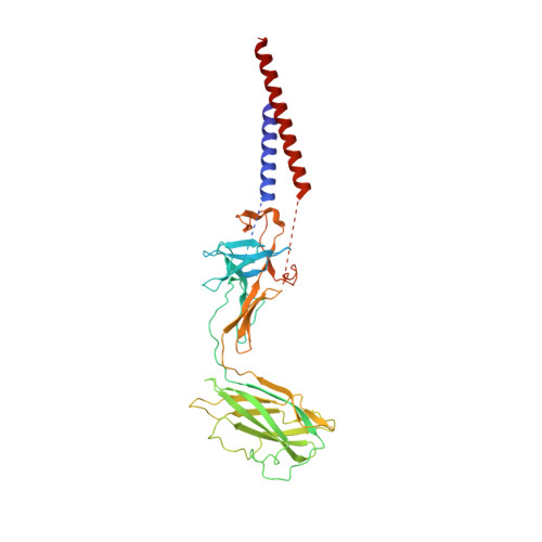
    The bacterial flagellar hook is a short, highly curved tubular structure connecting the rotary motor to the filament acting as a helical propeller. The bending flexibility of the hook allows it to work as a universal joint. A partial atomic model of the hook revealed a sliding intersubunit domain interaction along the protofilament to produce bending flexibility. However, it remained unclear how the tightly packed inner core domains can still permit axial extension and compression. We report advances in cryoEM image analysis for high-resolution, high-throughput structural analysis and a density map of the hook that reveals most of the secondary structures, including the terminal alpha helices forming a coiled coil. The orientations and axial packing interactions of these two alpha helices are distinctly different from those of the filament, allowing them to have a room for axial compression and extension for bending flexibility without impairing the mechanical stability of the hook.


  • Organizational Affiliation
    • Dynamic NanoMachine Project, ICORP, JST, 1-3 Yamadaoka, Suita, Osaka 565-0871, Japan.

Macromolecule Content 

  • Total Structure Weight: 42.1 kDa 
  • Atom Count: 2,573 
  • Modeled Residue Count: 347 
  • Deposited Residue Count: 402 
  • Unique protein chains: 1

Macromolecules

Find similar proteins by:|  3D Structure
Entity ID: 1
MoleculeChains  Sequence LengthOrganismDetailsImage
Flagellar hook protein flgE402Salmonella enterica subsp. enterica serovar TyphimuriumMutation(s): 0 
UniProt
Find proteins for P0A1J1 (Salmonella typhimurium (strain LT2 / SGSC1412 / ATCC 700720))
Explore P0A1J1 
Go to UniProtKB:  P0A1J1
Entity Groups
Sequence Clusters30% Identity50% Identity70% Identity90% Identity95% Identity100% Identity
UniProt GroupP0A1J1
Sequence Annotations
Expand
Reference Sequence

Experimental Data & Validation

Experimental Data

  • Method: ELECTRON MICROSCOPY
  • Resolution: 7.10 Å
  • Aggregation State: FILAMENT 
  • Reconstruction Method: HELICAL 

Structure Validation

View Full Validation Report


Entry History 

Deposition Data

Revision History  (Full details and data files)

  • Version 1.0: 2009-12-15
    Type: Initial release
  • Version 1.1: 2011-07-13
    Changes: Version format compliance
  • Version 1.2: 2019-10-09
    Changes: Author supporting evidence, Data collection, Database references, Other
  • Version 1.3: 2024-03-13
    Changes: Data collection, Database references, Derived calculations