3A5Y | pdb_00003a5y

Crystal structure of GenX from Escherichia coli in complex with lysyladenylate analog


Experimental Data Snapshot

  • Method: X-RAY DIFFRACTION
  • Resolution: 1.90 Å
  • R-Value Free: 
    0.219 (Depositor), 0.219 (DCC) 
  • R-Value Work: 
    0.171 (Depositor), 0.171 (DCC) 
  • R-Value Observed: 
    0.171 (Depositor) 

Starting Model: experimental
View more details

wwPDB Validation 3D Report Full Report

Validation slider image for 3A5Y

Ligand Structure Quality Assessment 


This is version 1.3 of the entry. See complete history

Literature

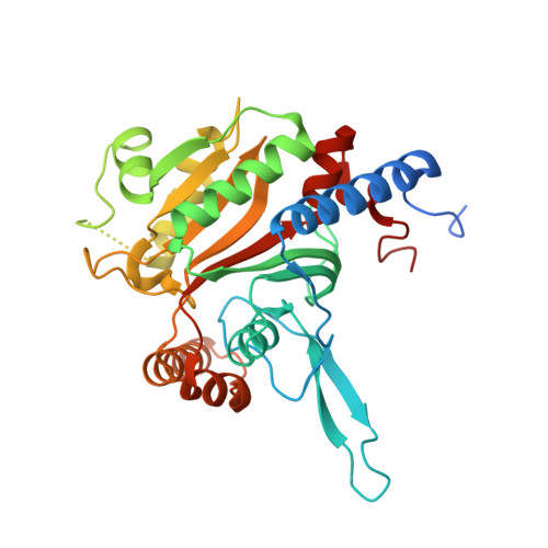
A paralog of lysyl-tRNA synthetase aminoacylates a conserved lysine residue in translation elongation factor P.

Yanagisawa, T.Sumida, T.Ishii, R.Takemoto, C.Yokoyama, S.

(2010) Nat Struct Mol Biol 17: 1136-1143

  • DOI: https://doi.org/10.1038/nsmb.1889
  • Primary Citation Related Structures: 
    3A5Y, 3A5Z

  • PubMed Abstract: 

    Aminoacyl-tRNA synthetase (aaRS) paralogs with unknown functions exist in various species. We now report novel 'protein lysylation' by an Escherichia coli lysyl-tRNA synthetase paralog, GenX/PoxA/YjeA. X-ray crystallographic analysis shows that the structure of the GenX protein resembles that of a class II aaRS. Further in vitro studies reveal that it specifically aminoacylates EF-P with lysine. The shape of the protein substrate mimics that of the L-shaped tRNA, and its lysylation site corresponds to the tRNA 3' end. Thus, we show how the aaRS architecture can be adapted to achieve aminoacylation of a specific protein. Moreover, in vivo analyses reveal that the translation elongation factor P (EF-P) lysylation by GenX is enhanced by YjeK (lysine 2,3-aminomutase paralog), which is encoded next to the EF-P gene, and might convert alpha-lysyl-EF-P to beta-lysyl-EF-P. In vivo analyses indicate that the EF-P modification by GenX and YjeK is essential for cell survival.


  • Organizational Affiliation
    • RIKEN Systems and Structural Biology Center, Tsurumi, Yokohama, Japan.

Macromolecule Content 

  • Total Structure Weight: 158.65 kDa 
  • Atom Count: 11,035 
  • Modeled Residue Count: 1,230 
  • Deposited Residue Count: 1,380 
  • Unique protein chains: 1

Macromolecules

Find similar proteins by:|  3D Structure
Entity ID: 1
MoleculeChains  Sequence LengthOrganismDetailsImage
Putative lysyl-tRNA synthetase
A, B, C, D
345Escherichia coliMutation(s): 0 
Gene Names: ECs5136GenX
EC: 6.1.1.6 (PDB Primary Data), 6.3.2 (UniProt)
UniProt
Find proteins for P0A8N7 (Escherichia coli (strain K12))
Explore P0A8N7 
Go to UniProtKB:  P0A8N7
Entity Groups
Sequence Clusters30% Identity50% Identity70% Identity90% Identity95% Identity100% Identity
UniProt GroupP0A8N7
Sequence Annotations
Expand
Reference Sequence

Experimental Data & Validation

Experimental Data

  • Method: X-RAY DIFFRACTION
  • Resolution: 1.90 Å
  • R-Value Free:  0.219 (Depositor), 0.219 (DCC) 
  • R-Value Work:  0.171 (Depositor), 0.171 (DCC) 
  • R-Value Observed: 0.171 (Depositor) 
Space Group: P 1
Unit Cell:
Length ( Å )Angle ( ˚ )
a = 54.8α = 95.47
b = 69.15β = 106.51
c = 94.08γ = 90.46
Software Package:
Software NamePurpose
HKL-2000data collection
MOLREPphasing
CNSrefinement
HKL-2000data reduction
SCALEPACKdata scaling

Structure Validation

View Full Validation Report


Ligand Structure Quality Assessment 


Entry History 

Deposition Data

Revision History  (Full details and data files)

  • Version 1.0: 2010-08-25
    Type: Initial release
  • Version 1.1: 2011-07-13
    Changes: Version format compliance
  • Version 1.2: 2013-10-23
    Changes: Database references
  • Version 1.3: 2023-11-01
    Changes: Data collection, Database references, Derived calculations, Refinement description