3A52 | pdb_00003a52

Crystal structure of cold-active alkailne phosphatase from psychrophile Shewanella sp.


Experimental Data Snapshot

  • Method: X-RAY DIFFRACTION
  • Resolution: 2.20 Å
  • R-Value Free: 
    0.244 (Depositor), 0.244 (DCC) 
  • R-Value Work: 
    0.183 (Depositor), 0.183 (DCC) 

wwPDB Validation 3D Report Full Report

Validation slider image for 3A52

This is version 1.4 of the entry. See complete history

Literature

Crystal structure of cold-active alkaline phosphatase from the psychrophile Shewanella sp.

Tsuruta, H.Mikami, B.Higashi, T.Aizono, Y.

(2010) Biosci Biotechnol Biochem 74: 69-74

  • DOI: https://doi.org/10.1271/bbb.90563
  • Primary Citation Related Structures: 
    3A52

  • PubMed Abstract: 

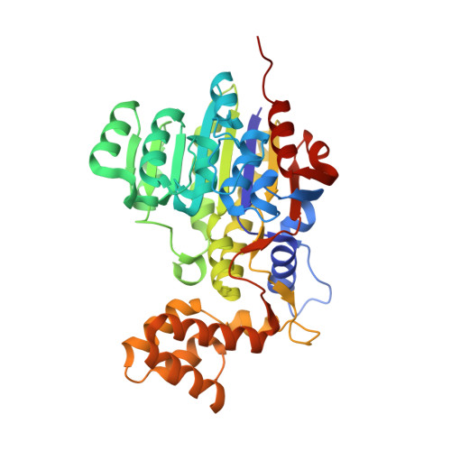
    The crystal structure of a cold-active alkaline phosphatase from a psychrophile, Shewanella sp. (SCAP), was solved at 2.2 A. A refined model showed a homodimer with six metal-ligand sites. The arrangement of the catalytic residues resembled those of alkaline phosphatases (APs), suggesting that the reaction mechanism of SCAP was fundamentally identical to those of other APs. SCAP had two distinct structural features: (i) a loop with Arg122 that bound to the phosphate moiety of the substrate suffered no constraints from the linkage to other secondary structures, and (ii) Mg3-ligand His109 was considered to undergo repulsive effect with neighboring Trp228. The local flexibility led by these features might be an important factor in the high catalytic efficiency of SCAP at low temperatures.


  • Organizational Affiliation
    • Office of Collaborative Research and Technology Development, Kobe University, Hyogo, Japan.

Macromolecule Content 

  • Total Structure Weight: 86.81 kDa 
  • Atom Count: 6,862 
  • Modeled Residue Count: 798 
  • Deposited Residue Count: 800 
  • Unique protein chains: 1

Macromolecules

Find similar proteins by:|  3D Structure
Entity ID: 1
MoleculeChains  Sequence LengthOrganismDetailsImage
Cold-active alkaline phosphatase
A, B
400Shewanella sp. AP1Mutation(s): 0 
Gene Names: scap
UniProt
Find proteins for Q8RQU7 (Shewanella sp. AP1)
Explore Q8RQU7 
Go to UniProtKB:  Q8RQU7
Entity Groups
Sequence Clusters30% Identity50% Identity70% Identity90% Identity95% Identity100% Identity
UniProt GroupQ8RQU7
Sequence Annotations
Expand
Reference Sequence

Small Molecules

Ligands 3 Unique
IDChains Name / Formula / InChI Key2D Diagram3D Interactions
SO4

Query on SO4


Download:Ideal Coordinates CCD File
C [auth A],
G [auth B],
H [auth B]
SULFATE ION
O4 S
QAOWNCQODCNURD-UHFFFAOYSA-L
ZN

Query on ZN


Download:Ideal Coordinates CCD File
D [auth A],
I [auth B]
ZINC ION
Zn
PTFCDOFLOPIGGS-UHFFFAOYSA-N
MG

Query on MG


Download:Ideal Coordinates CCD File
E [auth A],
F [auth A],
J [auth B],
K [auth B]
MAGNESIUM ION
Mg
JLVVSXFLKOJNIY-UHFFFAOYSA-N

Experimental Data & Validation

Experimental Data

  • Method: X-RAY DIFFRACTION
  • Resolution: 2.20 Å
  • R-Value Free:  0.244 (Depositor), 0.244 (DCC) 
  • R-Value Work:  0.183 (Depositor), 0.183 (DCC) 
Space Group: P 1 21 1
Unit Cell:
Length ( Å )Angle ( ˚ )
a = 64.059α = 90
b = 95.863β = 101.62
c = 71.058γ = 90
Software Package:
Software NamePurpose
DENZOdata reduction
SCALEPACKdata scaling
CNSrefinement
PDB_EXTRACTdata extraction
HKL-2000data collection
HKL-2000data reduction
CNSphasing

Structure Validation

View Full Validation Report


Entry History 

Deposition Data

Revision History  (Full details and data files)

  • Version 1.0: 2010-04-14
    Type: Initial release
  • Version 1.1: 2011-07-13
    Changes: Version format compliance
  • Version 1.2: 2017-10-11
    Changes: Refinement description
  • Version 1.3: 2019-10-09
    Changes: Data collection, Database references
  • Version 1.4: 2024-03-13
    Changes: Data collection, Database references, Derived calculations