3A4I | pdb_00003a4i

Crystal structure of GMP synthetase PH1347 from Pyrococcus horikoshii OT3


Experimental Data Snapshot

  • Method: X-RAY DIFFRACTION
  • Resolution: 1.79 Å
  • R-Value Free: 
    0.246 (Depositor), 0.248 (DCC) 
  • R-Value Work: 
    0.210 (Depositor), 0.209 (DCC) 
  • R-Value Observed: 
    0.212 (Depositor) 

wwPDB Validation 3D Report Full Report

Validation slider image for 3A4I

This is version 1.2 of the entry. See complete history

Literature

Crystal structure of the ATPPase subunit and its substrate-dependent association with the GATase Subunit: a novel regulatory mechanism for a two-subunit-type GMP synthetase from Pyrococcus horikoshii OT3.

Maruoka, S.Horita, S.Lee, W.C.Nagata, K.Tanokura, M.

(2010) J Mol Biology 395: 417-429

  • DOI: https://doi.org/10.1016/j.jmb.2009.10.053
  • Primary Citation Related Structures: 
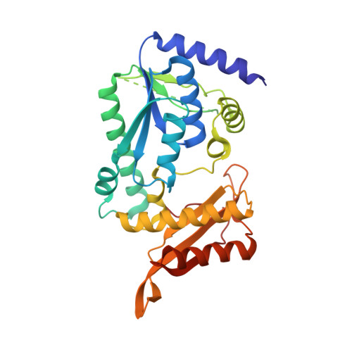
    3A4I

  • PubMed Abstract: 

    Guanosine 5'-monophosphate synthetase(s) (GMPS) catalyzes the final step of the de novo synthetic pathway of purine nucleotides. GMPS consists of two functional units that are present as domains or subunits: glutamine amidotransferase (GATase) and ATP pyrophosphatase (ATPPase). GATase hydrolyzes glutamine to yield glutamate and ammonia, while ATPPase utilizes ammonia to convert adenyl xanthosine 5'-monophosphate (adenyl-XMP) into guanosine 5'-monophosphate. Here we report the crystal structure of PH-ATPPase (the ATPPase subunit of the two-subunit-type GMPS from the hyperthermophilic archaeon Pyrococcus horikoshii OT3). PH-ATPPase consists of two domains (N-domain and C-domain) and exists as a homodimer in the crystal and in solution. The N-domain contains an ATP-binding platform called P-loop, whereas the C-domain contains the xanthosine 5'-monophosphate (XMP)-binding site and also contributes to homodimerization. We have also demonstrated that PH-GATase (the glutamine amidotransferase subunit of the two-subunit-type GMPS from the hyperthermophilic archaeon P. horikoshii OT3) alone is inactive, and that all substrates of PH-ATPPase except for ammonia (Mg(2+), ATP and XMP) are required to stabilize the active complex of PH-ATPPase and PH-GATase subunits.


  • Organizational Affiliation
    • Department of Applied Biological Chemistry, Graduate School of Agricultural and Life Sciences, University of Tokyo, 1-1-1 Yayoi, Bunkyo-ku, Tokyo 113-8657, Japan.

Macromolecule Content 

  • Total Structure Weight: 69.22 kDa 
  • Atom Count: 4,804 
  • Modeled Residue Count: 572 
  • Deposited Residue Count: 616 
  • Unique protein chains: 1

Macromolecules

Find similar proteins by:|  3D Structure
Entity ID: 1
MoleculeChains  Sequence LengthOrganismDetailsImage
GMP synthase [glutamine-hydrolyzing] subunit B
A, B
308Pyrococcus horikoshiiMutation(s): 0 
Gene Names: guaABPH1347
EC: 6.3.5.2
UniProt
Find proteins for O59072 (Pyrococcus horikoshii (strain ATCC 700860 / DSM 12428 / JCM 9974 / NBRC 100139 / OT-3))
Explore O59072 
Go to UniProtKB:  O59072
Entity Groups
Sequence Clusters30% Identity50% Identity70% Identity90% Identity95% Identity100% Identity
UniProt GroupO59072
Sequence Annotations
Expand
Reference Sequence

Experimental Data & Validation

Experimental Data

  • Method: X-RAY DIFFRACTION
  • Resolution: 1.79 Å
  • R-Value Free:  0.246 (Depositor), 0.248 (DCC) 
  • R-Value Work:  0.210 (Depositor), 0.209 (DCC) 
  • R-Value Observed: 0.212 (Depositor) 
Space Group: P 21 21 21
Unit Cell:
Length ( Å )Angle ( ˚ )
a = 69.903α = 90
b = 82.212β = 90
c = 111.565γ = 90
Software Package:
Software NamePurpose
REFMACrefinement
PDB_EXTRACTdata extraction
HKL-2000data collection
HKL-2000data reduction
HKL-2000data scaling
MOLREPphasing

Structure Validation

View Full Validation Report


Entry History 

Deposition Data

Revision History  (Full details and data files)

  • Version 1.0: 2009-07-21
    Type: Initial release
  • Version 1.1: 2011-07-13
    Changes: Advisory, Source and taxonomy, Version format compliance
  • Version 1.2: 2024-03-13
    Changes: Data collection, Database references