3A43 | pdb_00003a43

Crystal structure of HypA


Experimental Data Snapshot

  • Method: X-RAY DIFFRACTION
  • Resolution: 2.30 Å
  • R-Value Free: 
    0.239 (Depositor), 0.239 (DCC) 
  • R-Value Work: 
    0.215 (Depositor), 0.215 (DCC) 
  • R-Value Observed: 
    0.215 (Depositor) 

wwPDB Validation 3D Report Full Report

Validation slider image for 3A43

This is version 1.4 of the entry. See complete history

Literature

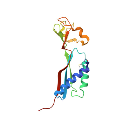
Crystal structure of HypA, a nickel-binding metallochaperone for [NiFe] hydrogenase maturation.

Watanabe, S.Arai, T.Matsumi, R.Atomi, H.Imanaka, T.Miki, K.

(2009) J Mol Biology 394: 448-459

  • DOI: https://doi.org/10.1016/j.jmb.2009.09.030
  • Primary Citation Related Structures: 
    3A43, 3A44

  • PubMed Abstract: 

    HypA is one of the auxiliary proteins involved in the maturation of [NiFe] hydrogenases. By an unknown mechanism, HypA functions as a metallochaperone in the insertion of the Ni atom into hydrogenases. We have determined the crystal structures of HypA from Thermococcus kodakaraensis KOD1 in both monomeric and dimeric states. The structure of the HypA monomer consists of Ni- and Zn-binding domains. The relative arrangement of the two metal-binding domains has been shown to be associated with local conformations of the conserved Ni-binding motif, suggesting a communication between the Ni- and Zn-binding sites. The HypA dimer has been shown to be stabilized by unexpected domain swapping through archaea-specific linker helices. In addition, the hexameric structure of HypA is formed in the crystal packing. Several hydrogen bonds and hydrophobic interactions stabilize the hexamer interface. These findings suggest the functional diversity of HypA proteins.


  • Organizational Affiliation
    • Department of Chemistry, Graduate School of Science, Kyoto University, Sakyo-ku, Kyoto, Japan.

Macromolecule Content 

  • Total Structure Weight: 31.61 kDa 
  • Atom Count: 2,110 
  • Modeled Residue Count: 257 
  • Deposited Residue Count: 278 
  • Unique protein chains: 1

Macromolecules

Find similar proteins by:|  3D Structure
Entity ID: 1
MoleculeChains  Sequence LengthOrganismDetailsImage
Hydrogenase nickel incorporation protein hypA
A, B
139Thermococcus kodakarensis KOD1Mutation(s): 0 
Gene Names: hypA
UniProt
Find proteins for Q5JIH3 (Thermococcus kodakarensis (strain ATCC BAA-918 / JCM 12380 / KOD1))
Explore Q5JIH3 
Go to UniProtKB:  Q5JIH3
Entity Groups
Sequence Clusters30% Identity50% Identity70% Identity90% Identity95% Identity100% Identity
UniProt GroupQ5JIH3
Sequence Annotations
Expand
Reference Sequence

Small Molecules

Modified Residues  1 Unique
IDChains TypeFormula2D DiagramParent
FME
Query on FME
A, B
L-PEPTIDE LINKINGC6 H11 N O3 SMET

Experimental Data & Validation

Experimental Data

  • Method: X-RAY DIFFRACTION
  • Resolution: 2.30 Å
  • R-Value Free:  0.239 (Depositor), 0.239 (DCC) 
  • R-Value Work:  0.215 (Depositor), 0.215 (DCC) 
  • R-Value Observed: 0.215 (Depositor) 
Space Group: H 3 2
Unit Cell:
Length ( Å )Angle ( ˚ )
a = 83.933α = 90
b = 83.933β = 90
c = 365.006γ = 120
Software Package:
Software NamePurpose
CNSrefinement
ADSCdata collection
HKL-2000data reduction
HKL-2000data scaling
SOLVEphasing

Structure Validation

View Full Validation Report


Entry History 

Deposition Data

Revision History  (Full details and data files)

  • Version 1.0: 2009-10-06
    Type: Initial release
  • Version 1.1: 2011-07-13
    Changes: Version format compliance
  • Version 1.2: 2014-01-01
    Changes: Database references, Other
  • Version 1.3: 2016-12-21
    Changes: Structure summary
  • Version 1.4: 2024-10-16
    Changes: Data collection, Database references, Derived calculations, Structure summary