3A40 | pdb_00003a40

Crystal structure of the human VDR ligand binding domain bound to the synthetic agonist compound 2alpha-methyl-AMCR277B(C23R)


Experimental Data Snapshot

  • Method: X-RAY DIFFRACTION
  • Resolution: 1.45 Å
  • R-Value Free: 
    0.196 (Depositor), 0.195 (DCC) 
  • R-Value Work: 
    0.160 (Depositor), 0.160 (DCC) 
  • R-Value Observed: 
    0.162 (Depositor) 

Starting Model: experimental
View more details

wwPDB Validation 3D Report Full Report

Validation slider image for 3A40

Ligand Structure Quality Assessment 


This is version 1.3 of the entry. See complete history

Literature

Structure-function relationships and crystal structures of the vitamin D receptor bound 2 alpha-methyl-(20S,23S)- and 2 alpha-methyl-(20S,23R)-epoxymethano-1 alpha,25-dihydroxyvitamin D3

Antony, P.Sigueiro, R.Huet, T.Sato, Y.Ramalanjaona, N.Rodrigues, L.C.Mourino, A.Moras, D.Rochel, N.

(2010) J Med Chem 53: 1159-1171

  • DOI: https://doi.org/10.1021/jm9014636
  • Primary Citation Related Structures: 
    3A3Z, 3A40

  • PubMed Abstract: 

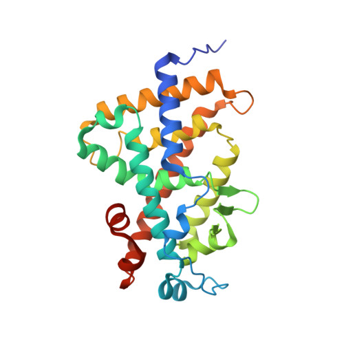
    The vitamin D nuclear receptor is a ligand-dependent transcription factor that controls multiple biological responses such as cell proliferation, immune responses, and bone mineralization. Numerous 1 alpha,25(OH)(2)D(3) analogues, which exhibit low calcemic side effects and/or antitumoral properties, have been synthesized. We recently showed that the synthetic analogue (20S,23S)-epoxymethano-1 alpha,25-dihydroxyvitamin D(3) (2a) acts as a 1 alpha,25(OH)(2)D(3) superagonist and exhibits both antiproliferative and prodifferentiating properties in vitro. Using this information and on the basis of the crystal structures of human VDR ligand binding domain (hVDR LBD) bound to 1 alpha,25(OH)(2)D(3), 2 alpha-methyl-1 alpha,25(OH)(2)D(3), or 2a, we designed a novel analogue, 2 alpha-methyl-(20S,23S)-epoxymethano-1 alpha,25-dihydroxyvitamin D(3) (4a), in order to increase its transactivation potency. Here, we solved the crystal structures of the hVDR LBD in complex with the 4a (C23S) and its epimer 4b (C23R) and determined their correlation with specific biological outcomes.


  • Organizational Affiliation
    • Departement de Biologie et de Génomique Structurales, Centre National de la Recherche Scientifique, Institut National de la Santé de la Recherche Médicale, Université de Strasbourg, 1 Rue Laurent Fries, 67404 Illkirch, France.

Macromolecule Content 

  • Total Structure Weight: 30.46 kDa 
  • Atom Count: 2,567 
  • Modeled Residue Count: 255 
  • Deposited Residue Count: 263 
  • Unique protein chains: 1

Macromolecules

Find similar proteins by:|  3D Structure
Entity ID: 1
MoleculeChains  Sequence LengthOrganismDetailsImage
Vitamin D3 receptorA [auth X]263Homo sapiensMutation(s): 0 
UniProt & NIH Common Fund Data Resources
Find proteins for P11473 (Homo sapiens)
Explore P11473 
Go to UniProtKB:  P11473
PHAROS:  P11473
GTEx:  ENSG00000111424 
Entity Groups
Sequence Clusters30% Identity50% Identity70% Identity90% Identity95% Identity100% Identity
UniProt GroupP11473
Sequence Annotations
Expand
Reference Sequence

Small Molecules

Ligands 2 Unique
IDChains Name / Formula / InChI Key2D Diagram3D Interactions
23R

Query on 23R


Download:Ideal Coordinates CCD File
B [auth X](1S,2S,3R,5Z,7E,14beta,17alpha,23R)-23-(2-hydroxy-2-methylpropyl)-2-methyl-20,24-epoxy-9,10-secochola-5,7,10-triene-1,3-diol
C29 H46 O4
CDTXSCLKUNDXBZ-MWPSFRNWSA-N
SO4

Query on SO4


Download:Ideal Coordinates CCD File
C [auth X],
D [auth X]
SULFATE ION
O4 S
QAOWNCQODCNURD-UHFFFAOYSA-L

Experimental Data & Validation

Experimental Data

  • Method: X-RAY DIFFRACTION
  • Resolution: 1.45 Å
  • R-Value Free:  0.196 (Depositor), 0.195 (DCC) 
  • R-Value Work:  0.160 (Depositor), 0.160 (DCC) 
  • R-Value Observed: 0.162 (Depositor) 
Space Group: P 21 21 21
Unit Cell:
Length ( Å )Angle ( ˚ )
a = 44.969α = 90
b = 51.496β = 90
c = 131.91γ = 90
Software Package:
Software NamePurpose
REFMACrefinement
MxCuBEdata collection
HKL-2000data reduction
HKL-2000data scaling
REFMACphasing

Structure Validation

View Full Validation Report


Ligand Structure Quality Assessment 


Entry History 

Deposition Data

Revision History  (Full details and data files)

  • Version 1.0: 2010-02-02
    Type: Initial release
  • Version 1.1: 2011-07-13
    Changes: Version format compliance
  • Version 1.2: 2013-11-20
    Changes: Database references
  • Version 1.3: 2023-11-01
    Changes: Data collection, Database references, Derived calculations, Refinement description, Structure summary