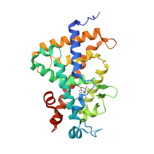
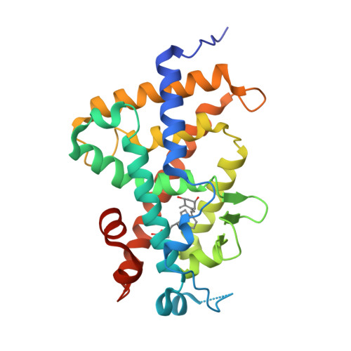
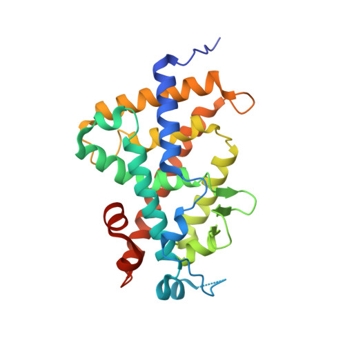
3A2I

Crystal structure of the human vitamin D receptor (H305F) ligand binding domain complexed with TEI-9647


Experimental Data Snapshot

  • Method: X-RAY DIFFRACTION
  • Resolution: 3.27 Å
  • R-Value Free: 0.276 
  • R-Value Work: 0.218 
  • R-Value Observed: 0.221 

Starting Model: experimental
View more details

wwPDB Validation   3D Report Full Report


Ligand Structure Quality Assessment 


This is version 1.3 of the entry. See complete history


Literature

Structural basis of the histidine-mediated vitamin D receptor agonistic and antagonistic mechanisms of (23S)-25-dehydro-1alpha-hydroxyvitamin D(3)-26,23-lactone

Kakuda, S.Ishizuka, S.Eguchi, H.Mizwicki, M.T.Norman, A.W.Takimoto-Kamimura, M.

(2010) Acta Crystallogr D Biol Crystallogr 66: 918-926

  • DOI: https://doi.org/10.1107/S0907444910020810
  • Primary Citation of Related Structures:  
    3A2H, 3A2I, 3A2J

  • PubMed Abstract: 

    TEI-9647 antagonizes vitamin D receptor (VDR) mediated genomic actions of 1alpha,25(OH)2D3 in human cells but is agonistic in rodent cells. The presence of Cys403, Cys410 or of both residues in the C-terminal region of human VDR (hVDR) results in antagonistic action of this compound. In the complexes of TEI-9647 with wild-type hVDR (hVDRwt) and H397F hVDR, TEI-9647 functions as an antagonist and forms a covalent adduct with hVDR according to MALDI-TOF MS. The crystal structures of complexes of TEI-9647 with rat VDR (rVDR), H305F hVDR and H305F/H397F hVDR showed that the agonistic activity of TEI-9647 is caused by a hydrogen-bond interaction with His397 or Phe397 located in helix 11. Both biological activity assays and the crystal structure of H305F hVDR complexed with TEI-9647 showed that the interaction between His305 and TEI-9647 is crucial for antagonist activity. This study indicates the following stepwise mechanism for TEI-9647 antagonism. Firstly, TEI-9647 forms hydrogen bonds to His305, which promote conformational changes in hVDR and draw Cys403 or Cys410 towards the ligand. This is followed by the formation of a 1,4-Michael addition adduct between the thiol (-SH) group of Cys403 or Cys410 and the exo-methylene group of TEI-9647.


  • Organizational Affiliation

    Teijin Institute for Bio-Medical Research, Japan.


Macromolecules
Find similar proteins by:  (by identity cutoff)  |  3D Structure
Entity ID: 1
MoleculeChains Sequence LengthOrganismDetailsImage
Vitamin D3 receptor263Homo sapiensMutation(s): 1 
Gene Names: VDRNR1I1
UniProt & NIH Common Fund Data Resources
Find proteins for P11473 (Homo sapiens)
Explore P11473 
Go to UniProtKB:  P11473
PHAROS:  P11473
GTEx:  ENSG00000111424 
Entity Groups  
Sequence Clusters30% Identity50% Identity70% Identity90% Identity95% Identity100% Identity
UniProt GroupP11473
Sequence Annotations
Expand
  • Reference Sequence
Small Molecules
Ligands 1 Unique
IDChains Name / Formula / InChI Key2D Diagram3D Interactions
TEJ
Query on TEJ

Download Ideal Coordinates CCD File 
B [auth A](1S,3R,5Z,7E,20S,23S)-1,3-dihydroxy-23,26-epoxy-9,10-secocholesta-5,7,10,25(27)-tetraen-26-one
C27 H38 O4
SAODSJHDCZTVAT-HPNZEMCUSA-N
Experimental Data & Validation

Experimental Data

  • Method: X-RAY DIFFRACTION
  • Resolution: 3.27 Å
  • R-Value Free: 0.276 
  • R-Value Work: 0.218 
  • R-Value Observed: 0.221 
  • Space Group: P 21 21 21
Unit Cell:
Length ( Å )Angle ( ˚ )
a = 44.796α = 90
b = 51.365β = 90
c = 131.773γ = 90
Software Package:
Software NamePurpose
REFMACrefinement
HKL-2000data collection
HKL-2000data reduction
HKL-2000data scaling
MOLREPphasing

Structure Validation

View Full Validation Report


Ligand Structure Quality Assessment 


Entry History 

Deposition Data

Revision History  (Full details and data files)

  • Version 1.0: 2010-05-26
    Type: Initial release
  • Version 1.1: 2011-07-13
    Changes: Version format compliance
  • Version 1.2: 2017-08-09
    Changes: Source and taxonomy
  • Version 1.3: 2023-11-01
    Changes: Data collection, Database references, Derived calculations, Refinement description