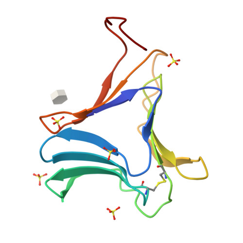
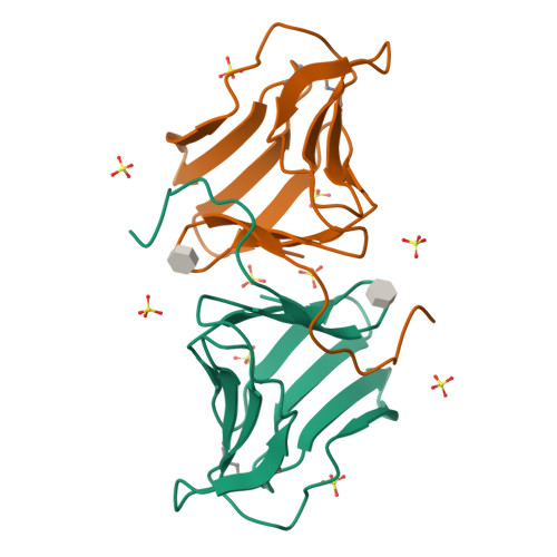
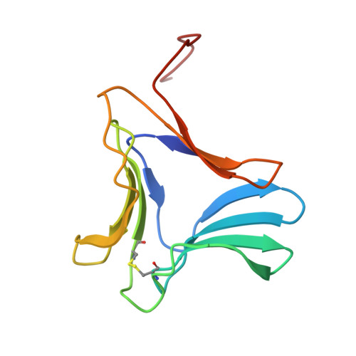
3A0D

Crystal Structure of Polygonatum cyrtonema lectin (PCL) complexed with monomannoside


Experimental Data Snapshot

  • Method: X-RAY DIFFRACTION
  • Resolution: 2.20 Å
  • R-Value Free: 0.217 
  • R-Value Work: 0.198 
  • R-Value Observed: 0.198 

Starting Model: experimental
View more details

wwPDB Validation   3D Report Full Report


Ligand Structure Quality Assessment 

Created with Raphaël 2.3.0Worse 01 BetterLigand structure goodness of fit to experimental dataBest fitted MMAClick on this verticalbar to view details

This is version 1.5 of the entry. See complete history


Literature

Crystal structures of a novel anti-HIV mannose-binding lectin from Polygonatum cyrtonema Hua with unique ligand-binding property and super-structure

Ding, J.Bao, J.Zhu, D.Zhang, Y.Wang, D.C.

(2010) J Struct Biol 171: 309-317

  • DOI: https://doi.org/10.1016/j.jsb.2010.05.009
  • Primary Citation of Related Structures:  
    3A0C, 3A0D, 3A0E

  • PubMed Abstract: 

    Polygonatum cyrtonema lectin (PCL) is a novel anti-HIV mannose-binding lectin from Galanthus nivalis agglutinin (GNA)-related lectin family. Crystal structures of ligand-free PCL and its complexes with monomannoside and alpha1-3 dimannoside have been determined. The ligand-free PCL is dimeric, with both subunits adopt the beta-prism II fold. PCL subunit binds mannose using a potential bivalent mode instead of the usual trivalent mode, in which carbohydrate-binding site (CBS) I and CBS III adopt the conserved mannose-binding motif of QXDXNXVXY (X is one of any amino acid residues) as observed in other structurally characterized GNA-related lectins, while CBS II adopts a modified motif with residues Gln58 and Asp60, which are critical for mannose-binding, substituted by His58 and Asn60, respectively. As a result, CBS II is unfit for mannose-binding. In the mannoside complexes, ligand-bindings only occur at CBS I which provides the specificity for alpha1-3 dimannoside. CBS II and CBS III are cooperatively occupied by a well-ordered sulfate ion, through which the individual dimers are cross-linked to form a unique super-structure of 3(2) helical lattice. Surveying the sequences of GNA-related lectins revealed that the modified binding motif of CBS II is widely distributed in the Liliaceae family as an intrinsic structural element. There is evidence that other GNA-related lectins will also adopt the similar super-structure as PCL. Thus PCL structure, unique in ligand-binding mode, may represent a novel type of structure of GNA-related lectins. Comparative analyses indicated that the dimer-based super-structure may play a primary role in the anti-HIV property of PCL.


  • Organizational Affiliation

    National Laboratory of Biomacromolecules, Institute of Biophysics, Chinese Academy of Sciences, Beijing 100101, People's Republic of China.


Macromolecules
Find similar proteins by:  (by identity cutoff)  |  3D Structure
Entity ID: 1
MoleculeChains Sequence LengthOrganismDetailsImage
Mannose/sialic acid-binding lectin110Polygonatum cyrtonemaMutation(s): 0 
UniProt
Find proteins for Q8L568 (Polygonatum cyrtonema)
Explore Q8L568 
Go to UniProtKB:  Q8L568
Entity Groups  
Sequence Clusters30% Identity50% Identity70% Identity90% Identity95% Identity100% Identity
UniProt GroupQ8L568
Sequence Annotations
Expand
  • Reference Sequence
Experimental Data & Validation

Experimental Data

  • Method: X-RAY DIFFRACTION
  • Resolution: 2.20 Å
  • R-Value Free: 0.217 
  • R-Value Work: 0.198 
  • R-Value Observed: 0.198 
  • Space Group: P 32 2 1
Unit Cell:
Length ( Å )Angle ( ˚ )
a = 76.46α = 90
b = 76.46β = 90
c = 61.75γ = 120
Software Package:
Software NamePurpose
CrystalCleardata collection
PHASERphasing
CNSrefinement
MOSFLMdata reduction
SCALAdata scaling

Structure Validation

View Full Validation Report


Ligand Structure Quality Assessment 

Created with Raphaël 2.3.0Worse 01 BetterLigand structure goodness of fit to experimental dataBest fitted MMAClick on this verticalbar to view details

Entry History 

Deposition Data

Revision History  (Full details and data files)

  • Version 1.0: 2010-03-16
    Type: Initial release
  • Version 1.1: 2011-07-13
    Changes: Version format compliance
  • Version 1.2: 2012-03-21
    Changes: Database references
  • Version 1.3: 2020-07-29
    Type: Remediation
    Reason: Carbohydrate remediation
    Changes: Data collection, Derived calculations, Structure summary
  • Version 1.4: 2023-11-01
    Changes: Data collection, Database references, Refinement description, Structure summary
  • Version 1.5: 2024-11-20
    Changes: Structure summary