37US | pdb_000037us

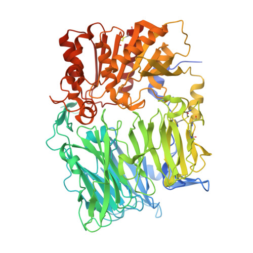
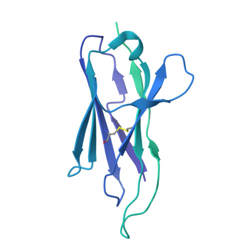
Cryo-EM structure of human Fibroblast Activation Protein alpha in complex with H4 VNAR


Experimental Data Snapshot

  • Method: ELECTRON MICROSCOPY
  • Resolution: 3.01 Å
  • Aggregation State: PARTICLE 
  • Reconstruction Method: SINGLE PARTICLE 

Starting Model: experimental
View more details

wwPDB Validation 3D Report Full Report

Validation slider image for 37US

This is version 1.0 of the entry. See complete history. 

Literature

A shark variable new antigen receptor recognizes an occluded epitope of fibroblast activation protein.

Broadberry, R.L., Ott, K.L., Lake, E.W., Ravi, T., Hemme, C.A., Chi, S., West, J.L., Gunaratne, G.S., Ong, I.M., LeBeau, A.M., Grant, T.

(2026) bioRxiv 

  • DOI: https://doi.org/10.64898/2026.08.11.744296
  • Primary Citation Related Structures: 
    37US, 37UT, 37UU

  • PubMed Abstract: 

    Variable new antigen receptors (VNARs) are the smallest naturally occurring antibody binding domains. Their size allows VNARs to access sterically restricted epitopes that are inaccessible to conventional antibodies. We recently identified a suite of VNARs that target fibroblast activation protein (FAP), a stromal serine protease indicative of extracellular matrix remodeling. The presence of FAP on the surface of cancer-associated fibroblasts (CAFs) that promote immunosuppression has made FAP a compelling therapeutic target for cancer therapy. Although antibodies targeting FAP have been developed, there is a paucity of information on how biologics engage FAP. Here, we used single-particle cryogenic electron microscopy (cryo-EM) to compare FAP recognition of three antibody architectures: a shark-derived VNAR, variable heavy (V H ) and light domains (V L ) of a humanized Immunoglobulin G (IgG), and a camelid-derived VHH. The humanized V H -V L domains and camelid VHH both target a solvent-exposed β-propeller domain, whereas the VNAR binds a highly conserved, topologically recessed epitope at the FAP dimer interface. Radical-footprinting mass spectrometry (MS) further mapped two additional immune-derived VNARs to distinct FAP surfaces outside the shared β-propeller epitope. These findings demonstrate how unique VNAR architecture can expand access to underexplored FAP surfaces and establish a structural framework for rational multiepitope targeting strategies.


  • Organizational Affiliation: 
    • John and Jean Rowe Institute of Virology, Morgridge Institute for Research, Madison, WI 53715, USA.

Macromolecule Content 

  • Total Structure Weight: 254.55 kDa 
  • Atom Count: 13,686 
  • Modeled Residue Count: 1,656 
  • Deposited Residue Count: 2,198 
  • Unique protein chains: 2

Macromolecules

Find similar proteins by:|  3D Structure
Entity ID: 1
MoleculeChains  Sequence LengthOrganismDetailsImage
Fibroblast activation protein alphaA,
C [auth B]
753Homo sapiensMutation(s): 0 
Gene Names: FAP
EC: 3.4.14.5 (PDB Primary Data), 3.4.21 (PDB Primary Data), 3.4.21.26 (PDB Primary Data)
UniProt & NIH Common Fund Data Resources
Find proteins for Q12884 (Homo sapiens)
Explore Q12884 
Go to UniProtKB:  Q12884
PHAROS:  Q12884
GTEx:  ENSG00000078098 
Entity Groups
Sequence Clusters30% Identity50% Identity70% Identity90% Identity95% Identity100% Identity
UniProt GroupQ12884
Glycosylation
Glycosylation Sites: 6Go to GlyGen: Q12884-1
Sequence Annotations
Expand
Reference Sequence
Find similar proteins by:|  3D Structure
Entity ID: 2
MoleculeChains  Sequence LengthOrganismDetailsImage
H4 variable new antigen receptor-human IgG Fc fusion proteinB [auth C],
D
346Ginglymostoma cirratum, Homo sapiens
This entity is chimeric
Mutation(s): 0 
Gene Names: NAR
UniProt
Find proteins for Q8JGJ1 (Ginglymostoma cirratum)
Explore Q8JGJ1 
Go to UniProtKB:  Q8JGJ1
Find proteins for P0DOX5 (Homo sapiens)
Explore P0DOX5 
Go to UniProtKB:  P0DOX5
Entity Groups
Sequence Clusters30% Identity50% Identity70% Identity90% Identity95% Identity100% Identity
UniProt GroupsP0DOX5Q8JGJ1
Sequence Annotations
Expand
Reference Sequence

Oligosaccharides

Help  
Entity ID: 3
MoleculeChains Length2D Diagram GlycosylationD Interactions
2-acetamido-2-deoxy-beta-D-glucopyranose-(1-4)-2-acetamido-2-deoxy-beta-D-glucopyranose
E, F, G, H, I
E, F, G, H, I, J
2N-Glycosylation

Experimental Data & Validation

Experimental Data

  • Method: ELECTRON MICROSCOPY
  • Resolution: 3.01 Å
  • Aggregation State: PARTICLE 
  • Reconstruction Method: SINGLE PARTICLE 
EM Software:
TaskSoftware PackageVersion
RECONSTRUCTIONcisTEM
MODEL REFINEMENTPHENIX
MODEL REFINEMENTCoot

Structure Validation

View Full Validation Report


Entry History 

& Funding Information

Deposition Data


Funding OrganizationLocationGrant Number
National Institutes of Health/National Cancer Institute (NIH/NCI)United StatesR01 CA237272
National Institutes of Health/National Cancer Institute (NIH/NCI)United StatesR01 CA233562
National Institutes of Health/National Cancer Institute (NIH/NCI)United StatesR01 CA305895
National Institutes of Health/National Cancer Institute (NIH/NCI)United StatesR01 CA295576
National Institutes of Health/National Institute of General Medical Sciences (NIH/NIGMS)United StatesT32 GM130550

Revision History  (Full details and data files)

  • Version 1.0: 2026-11-04
    Type: Initial release