36WN | pdb_000036wn

Mevalonate kinase from Saccharomyces cerevisiae with geranylgeranyl pyrophosphate (GGPP) bound


Experimental Data Snapshot

  • Method: X-RAY DIFFRACTION
  • Resolution: 1.95 Å
  • R-Value Free: 
    0.228 (Depositor), 0.228 (DCC) 
  • R-Value Work: 
    0.203 (Depositor), 0.203 (DCC) 
  • R-Value Observed: 
    0.205 (Depositor) 

Starting Model: experimental
View more details

wwPDB Validation 3D Report Full Report

Validation slider image for 36WN

Ligand Structure Quality Assessment 


This is version 1.0 of the entry. See complete history. 

Literature

Structural basis of mevalonate pathway regulation by feedback inhibition of mevalonate kinase.

D'Emilia, R.L.S., McCaskey, K.A., Ragwan, E.R., Kim, J.H., Chang, V., Tang, M.M., Kung, Y.

(2026) J Biol Chem : 113566-113566

  • DOI: https://doi.org/10.1016/j.jbc.2026.113566
  • Primary Citation Related Structures: 
    36WI, 36WJ, 36WK, 36WL, 36WM, 36WN

  • PubMed Abstract: 

    The mevalonate pathway diverts acetyl-CoA from central metabolism to produce isopentenyl pyrophosphate (IPP) and dimethylallyl pyrophosphate (DMAPP), the universal precursors to all steroids and isoprenoids in biology. Mevalonate kinase (MK) catalyzes a key regulatory step of mevalonate pathway in many eukaryotes, in which several downstream isoprenoid pyrophosphates act as feedback inhibitors. Known MK inhibitors vary widely in size, including IPP and DMAPP (five carbons, C 5 ), geranyl pyrophosphate (GPP, C 10 ), farnesyl pyrophosphate (FPP, C 15 ), and geranylgeranyl pyrophosphate (GGPP, C 20 ). These inhibitors compete for binding at a single site, yet how the enzyme is able to accommodate inhibitors of such varying sizes remains unknown. In this work, we first characterized the inhibition kinetics of all five of these inhibitors using MK from Saccharomyces cerevisiae (ScMK), revealing an inhibition potency that ranges approximately two orders of magnitude. We then solved X-ray crystal structures of ScMK bound to all five inhibitors as well as the structure of apo ScMK to shed light on how inhibitors of such varying sizes may all bind to the same site. In all, this suite of inhibitor-bound MK structures provides the first comprehensive structural depiction of MK feedback inhibition, a key regulatory mechanism of the mevalonate pathway of steroid and isoprenoid precursor biosynthesis.


  • Organizational Affiliation: 
    • Department of Chemistry, Bryn Mawr College, Bryn Mawr, PA 19010.

Macromolecule Content 

  • Total Structure Weight: 52.39 kDa 
  • Atom Count: 3,639 
  • Modeled Residue Count: 452 
  • Deposited Residue Count: 468 
  • Unique protein chains: 1

Macromolecules

Find similar proteins by:|  3D Structure
Entity ID: 1
MoleculeChains  Sequence LengthOrganismDetailsImage
Mevalonate kinase468Saccharomyces cerevisiaeMutation(s): 0 
Gene Names: ERG12, RAR1, YMR208W, YM8261.02
EC: 2.7.1.36
UniProt
Find proteins for P07277 (Saccharomyces cerevisiae (strain ATCC 204508 / S288c))
Explore P07277 
Go to UniProtKB:  P07277
Entity Groups
Sequence Clusters30% Identity50% Identity70% Identity90% Identity95% Identity100% Identity
UniProt GroupP07277
Sequence Annotations
Expand
Reference Sequence

Small Molecules

Ligands 5 Unique
IDChains Name / Formula / InChI Key2D Diagram3D Interactions
GRG
(Subject of Investigation/LOI)

Query on GRG


Download:Ideal Coordinates CCD File
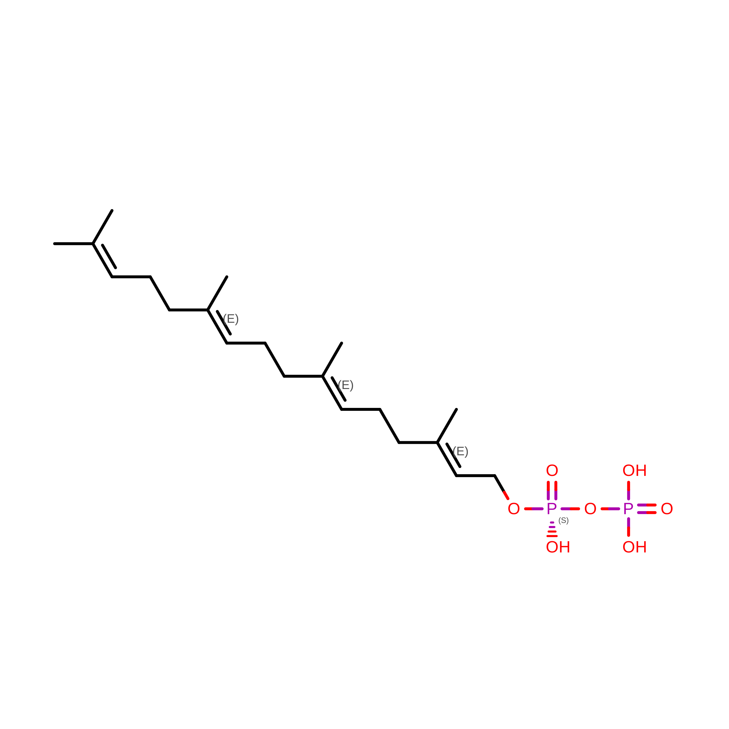
C [auth A]GERANYLGERANYL DIPHOSPHATE
C20 H36 O7 P2
OINNEUNVOZHBOX-QIRCYJPOSA-N
BGC

Query on BGC


Download:Ideal Coordinates CCD File
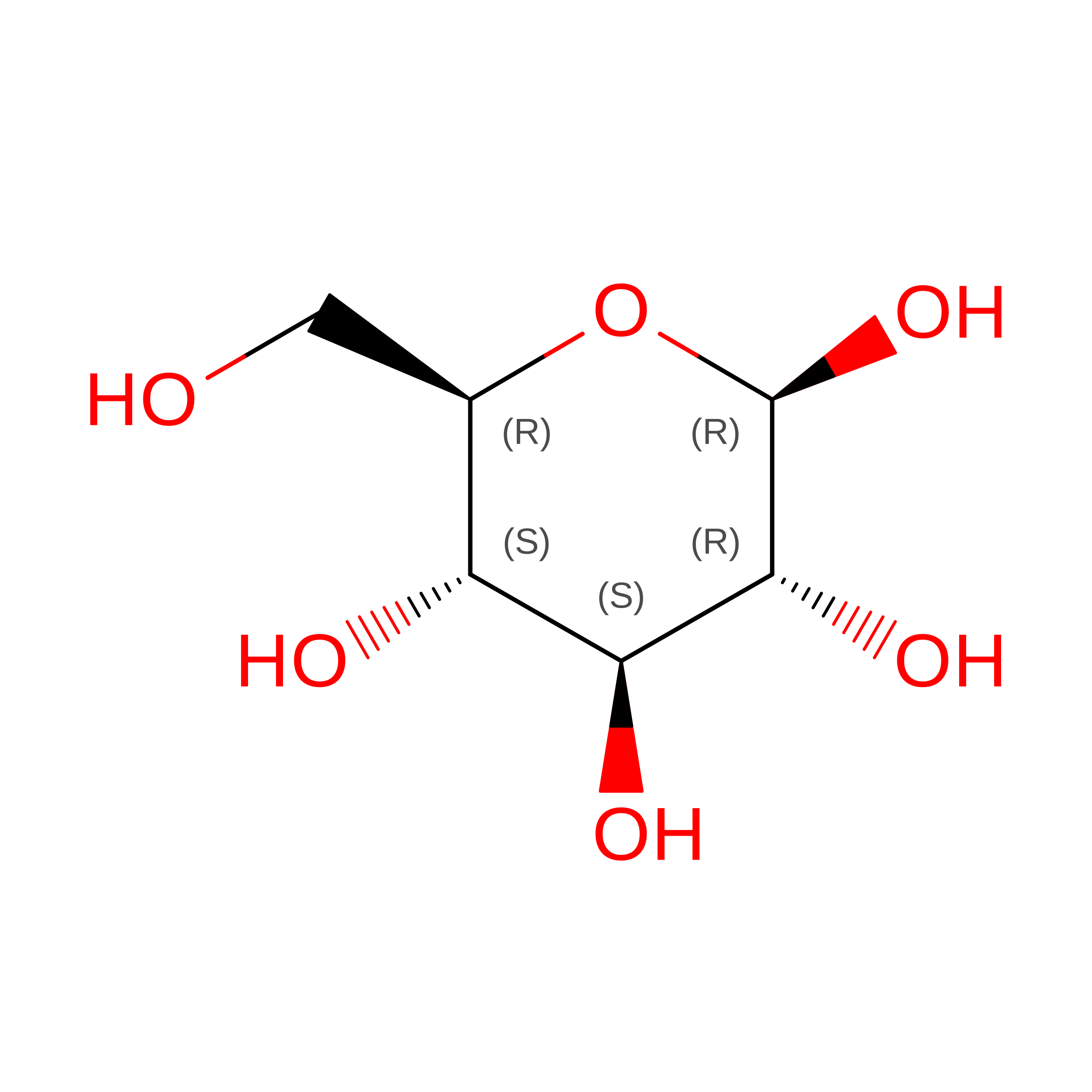
E [auth A]beta-D-glucopyranose
C6 H12 O6
WQZGKKKJIJFFOK-VFUOTHLCSA-N
TAR

Query on TAR


Download:Ideal Coordinates CCD File
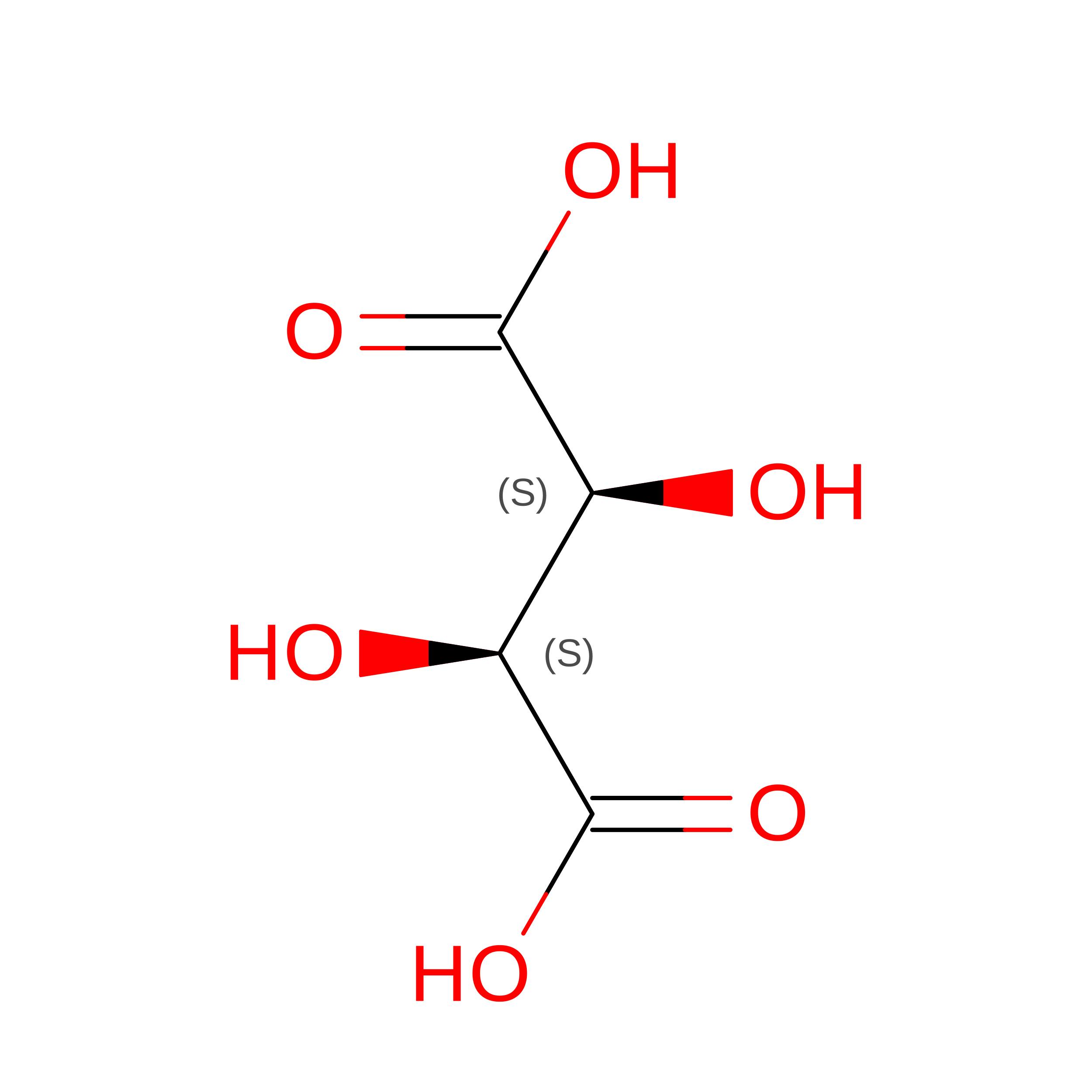
D [auth A]D(-)-TARTARIC ACID
C4 H6 O6
FEWJPZIEWOKRBE-LWMBPPNESA-N
GOL

Query on GOL


Download:Ideal Coordinates CCD File
F [auth A]GLYCEROL
C3 H8 O3
PEDCQBHIVMGVHV-UHFFFAOYSA-N
MG

Query on MG


Download:Ideal Coordinates CCD File
B [auth A]MAGNESIUM ION
Mg
JLVVSXFLKOJNIY-UHFFFAOYSA-N

Experimental Data & Validation

Experimental Data

  • Method: X-RAY DIFFRACTION
  • Resolution: 1.95 Å
  • R-Value Free:  0.228 (Depositor), 0.228 (DCC) 
  • R-Value Work:  0.203 (Depositor), 0.203 (DCC) 
  • R-Value Observed: 0.205 (Depositor) 
Space Group: P 43 21 2
Unit Cell:
Length ( Å )Angle ( ˚ )
a = 83.45α = 90
b = 83.45β = 90
c = 262.88γ = 90
Software Package:
Software NamePurpose
PHENIXrefinement
XDSdata reduction
XDSdata scaling
PHENIXphasing

Structure Validation

View Full Validation Report


Ligand Structure Quality Assessment 


Entry History 

& Funding Information

Deposition Data


Funding OrganizationLocationGrant Number
National Institutes of Health/National Institute of General Medical Sciences (NIH/NIGMS)United StatesGM116029

Revision History  (Full details and data files)

  • Version 1.0: 2026-09-30
    Type: Initial release