36CR | pdb_000036cr

Structure of the Evo-Phi36 bacteriophage


Experimental Data Snapshot

  • Method: ELECTRON MICROSCOPY
  • Resolution: 2.90 Å
  • Aggregation State: PARTICLE 
  • Reconstruction Method: SINGLE PARTICLE 

Starting Model: in silico
View more details

wwPDB Validation 3D Report Full Report

Validation slider image for 36CR

This is version 1.2 of the entry. See complete history

Literature

Generative design of bacteriophages with genome language models.

King, S.H.Driscoll, C.L.Li, D.B.Guo, D.Merchant, A.T.Brixi, G.Wilkinson, M.E.Hie, B.L.

(2026) Science 393: eaec2657-eaec2657

  • DOI: https://doi.org/10.1126/science.aec2657
  • Primary Citation Related Structures: 
    36CQ, 36CR

  • PubMed Abstract: 

    Many important biological functions arise not from single genes but from complex interactions encoded by entire genomes. We report the first generative design of complete bacteriophage genomes using genome language models. We generated viable bacteriophages with target host tropism, using the phage ΦX174 as our design template. Experimental testing yielded 16 phages with diverse fitness profiles in laboratory conditions. Cryo-electron microscopy confirmed that a generated phage utilizes an evolutionarily distant DNA packaging protein in its capsid. A cocktail of generated phages rapidly overcomes ΦX174-resistant Escherichia coli strains, demonstrating a path toward artificial intelligence-generated phage therapies against rapidly evolving bacterial pathogens. This work provides a blueprint for the design of diverse synthetic bacteriophages and useful biological systems at the genome scale.


  • Organizational Affiliation
    • Department of Bioengineering, Stanford University, Stanford, CA, USA.

Macromolecule Content 

  • Total Structure Weight: 70.41 kDa 
  • Atom Count: 4,849 
  • Modeled Residue Count: 612 
  • Deposited Residue Count: 627 
  • Unique protein chains: 3

Macromolecules

Find similar proteins by:  Sequence   |   3D Structure  
Entity ID: 1
MoleculeChains  Sequence LengthOrganismDetailsImage
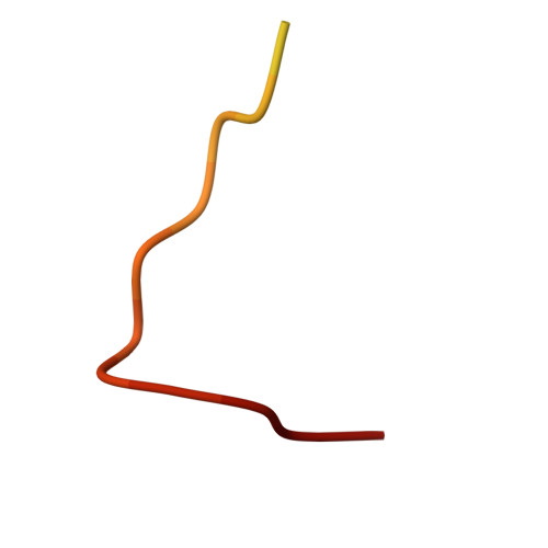
DNA-binding protein J25SinsheimervirusMutation(s): 0 
Entity Groups
Sequence Clusters30% Identity50% Identity70% Identity90% Identity95% Identity100% Identity
Sequence Annotations
Expand
Reference Sequence
Find similar proteins by:|  3D Structure
Entity ID: 2
MoleculeChains  Sequence LengthOrganismDetailsImage
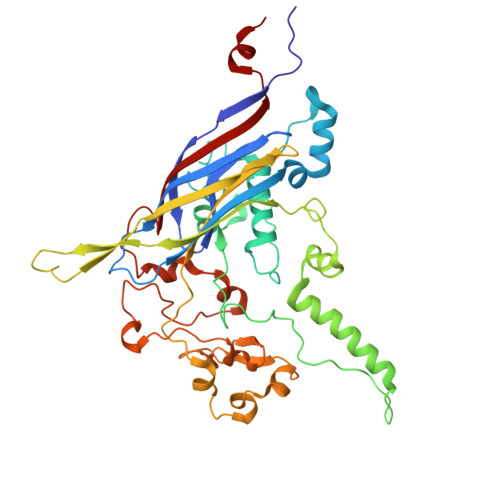
Capsid protein F427SinsheimervirusMutation(s): 0 
Entity Groups
Sequence Clusters30% Identity50% Identity70% Identity90% Identity95% Identity100% Identity
Sequence Annotations
Expand
Reference Sequence
Find similar proteins by:|  3D Structure
Entity ID: 3
MoleculeChains  Sequence LengthOrganismDetailsImage
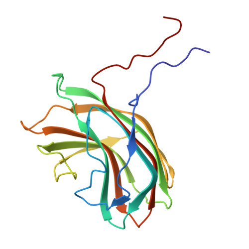
Major spike protein G175SinsheimervirusMutation(s): 0 
Entity Groups
Sequence Clusters30% Identity50% Identity70% Identity90% Identity95% Identity100% Identity
Sequence Annotations
Expand
Reference Sequence

Experimental Data & Validation

Experimental Data

  • Method: ELECTRON MICROSCOPY
  • Resolution: 2.90 Å
  • Aggregation State: PARTICLE 
  • Reconstruction Method: SINGLE PARTICLE 
EM Software:
TaskSoftware PackageVersion
RECONSTRUCTIONcryoSPARC4.6.2
RECONSTRUCTIONRELION5.0
MODEL REFINEMENTPHENIX2.0_5936

Structure Validation

View Full Validation Report


Entry History 

& Funding Information

Deposition Data


Funding OrganizationLocationGrant Number
Not funded--

Revision History  (Full details and data files)

  • Version 1.0: 2026-07-08
    Type: Initial release
  • Version 1.1: 2026-08-12
    Changes: Data collection, Database references
  • Version 1.2: 2026-08-19
    Changes: Data collection, Database references