34OC | pdb_000034oc

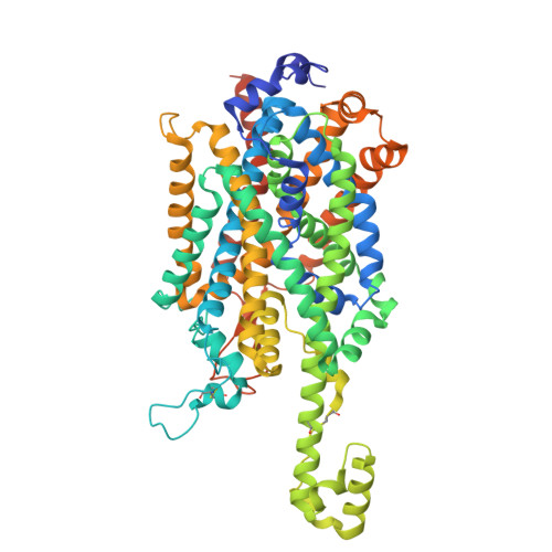
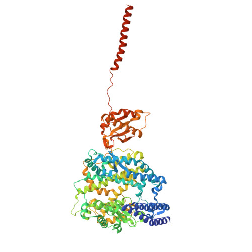
Structure of human apo B0AT1-ACE2 complex


Experimental Data Snapshot

  • Method: ELECTRON MICROSCOPY
  • Resolution: 3.19 Å
  • Aggregation State: PARTICLE 
  • Reconstruction Method: SINGLE PARTICLE 

Starting Model: experimental
View more details

wwPDB Validation 3D Report Full Report

Validation slider image for 34OC

This is version 1.0 of the entry. See complete history. 

Literature

Structural mechanisms of B 0 AT1 substrate recognition and complex assembly.

Pedersen, C.N., Christensen, M.E., Heger, T., Prakaash, D., Khalid, S., Shahsavar, A., Nissen, P., Stock, C.

(2026) J Mol Biol : 170086-170086

  • DOI: https://doi.org/10.1016/j.jmb.2026.170086
  • Primary Citation Related Structures: 
    34OA, 34OC, 34OD, 34OE, 34OH

  • PubMed Abstract: 

    Amino acid transporters are essential for maintaining cellular amino acid homeostasis. The broad-specificity neutral amino acid transporter B 0 AT1 is predominantly expressed in the intestine and kidney, where it mediates the uptake and reuptake of neutral amino acids with varying affinities. Proper plasma-membrane localization of B 0 AT1 requires dimerization with angiotensin-converting enzyme 2 (ACE2) in the intestine or collectrin in the kidney. Structural information is available for apo and substrate-bound B 0 AT1-ACE2 complexes, but how B 0 AT1 interacts with different accessory proteins and recognizes smaller substrates remains enigmatic. Here, we compare cryo-EM structures of B 0 AT1 in complex with ACE2 or collectrin in substrate-free states, and bound to alanine, leucine or tryptophan. The complexes display only minor differences in accessory-protein binding through a conserved interaction surface. Substrate recognition follows a canonical architecture observed for other SLC6 family transporters, with subtle adaptations that potentially account for the different affinities of the three amino acids. Notably, we resolve several cholesterol molecules surrounding the transporter, highlighting a potential regulatory role of the membrane environment that we address by molecular dynamics simulations.


  • Organizational Affiliation: 
    • Department of Molecular Biology and Genetics, Aarhus University, Aarhus, Denmark; DANDRITE, Nordic EMBL Partnership for Molecular Medicine, Denmark.

Macromolecule Content 

  • Total Structure Weight: 345.28 kDa 
  • Atom Count: 22,792 
  • Modeled Residue Count: 2,714 
  • Deposited Residue Count: 2,912 
  • Unique protein chains: 2

Macromolecules

Find similar proteins by:|  3D Structure
Entity ID: 1
MoleculeChains  Sequence LengthOrganismDetailsImage
Sodium-dependent neutral amino acid transporter B(0)AT1
A, B
643Homo sapiensMutation(s): 0 
Gene Names: SLC6A19, B0AT1
UniProt & NIH Common Fund Data Resources
Find proteins for Q695T7 (Homo sapiens)
Explore Q695T7 
Go to UniProtKB:  Q695T7
PHAROS:  Q695T7
GTEx:  ENSG00000174358 
Entity Groups
Sequence Clusters30% Identity50% Identity70% Identity90% Identity95% Identity100% Identity
UniProt GroupQ695T7
Glycosylation
Glycosylation Sites: 5Go to GlyGen: Q695T7-1
Sequence Annotations
Expand
Reference Sequence
Find similar proteins by:|  3D Structure
Entity ID: 2
MoleculeChains  Sequence LengthOrganismDetailsImage
Angiotensin-converting enzyme 2
C, D
813Homo sapiensMutation(s): 1 
Gene Names: ACE2, UNQ868/PRO1885
EC: 3.4.17.23 (PDB Primary Data), 3.4.17 (PDB Primary Data)
UniProt & NIH Common Fund Data Resources
Find proteins for Q9BYF1 (Homo sapiens)
Explore Q9BYF1 
Go to UniProtKB:  Q9BYF1
PHAROS:  Q9BYF1
GTEx:  ENSG00000130234 
Entity Groups
Sequence Clusters30% Identity50% Identity70% Identity90% Identity95% Identity100% Identity
UniProt GroupQ9BYF1
Glycosylation
Glycosylation Sites: 8Go to GlyGen: Q9BYF1-1
Sequence Annotations
Expand
Reference Sequence

Oligosaccharides

Help  
Entity ID: 3
MoleculeChains Length2D Diagram GlycosylationD Interactions
beta-D-mannopyranose-(1-4)-2-acetamido-2-deoxy-beta-D-glucopyranose-(1-4)-2-acetamido-2-deoxy-beta-D-glucopyranose
E, F, G, H, M
E, F, G, H, M, R
3N-Glycosylation
Entity ID: 4
MoleculeChains Length2D Diagram GlycosylationD Interactions
2-acetamido-2-deoxy-beta-D-glucopyranose-(1-4)-2-acetamido-2-deoxy-beta-D-glucopyranose
I, K, L, N, P
I, K, L, N, P, Q
2N-Glycosylation
Entity ID: 5
MoleculeChains Length2D Diagram GlycosylationD Interactions
2-acetamido-2-deoxy-beta-D-glucopyranose-(1-4)-[alpha-L-fucopyranose-(1-6)]2-acetamido-2-deoxy-beta-D-glucopyranose
J, O
3N-Glycosylation

Small Molecules

Ligands 4 Unique
IDChains Name / Formula / InChI Key2D Diagram3D Interactions
CLR

Query on CLR


Download:Ideal Coordinates CCD File
DA [auth B]
EA [auth B]
FA [auth B]
GA [auth B]
HA [auth B]
DA [auth B],
EA [auth B],
FA [auth B],
GA [auth B],
HA [auth B],
V [auth A],
W [auth A],
X [auth A],
Y [auth A],
Z [auth A]
CHOLESTEROL
C27 H46 O
HVYWMOMLDIMFJA-DPAQBDIFSA-N
A2G

Query on A2G


Download:Ideal Coordinates CCD File
KA [auth C],
OA [auth D]
2-acetamido-2-deoxy-alpha-D-galactopyranose
C8 H15 N O6
OVRNDRQMDRJTHS-CBQIKETKSA-N
NAG

Query on NAG


Download:Ideal Coordinates CCD File
AA [auth B]
BA [auth B]
CA [auth B]
IA [auth C]
JA [auth C]
AA [auth B],
BA [auth B],
CA [auth B],
IA [auth C],
JA [auth C],
MA [auth D],
NA [auth D],
S [auth A],
T [auth A],
U [auth A]
2-acetamido-2-deoxy-beta-D-glucopyranose
C8 H15 N O6
OVRNDRQMDRJTHS-FMDGEEDCSA-N
ZN

Query on ZN


Download:Ideal Coordinates CCD File
LA [auth C],
PA [auth D]
ZINC ION
Zn
PTFCDOFLOPIGGS-UHFFFAOYSA-N

Experimental Data & Validation

Experimental Data

  • Method: ELECTRON MICROSCOPY
  • Resolution: 3.19 Å
  • Aggregation State: PARTICLE 
  • Reconstruction Method: SINGLE PARTICLE 
EM Software:
TaskSoftware PackageVersion
RECONSTRUCTIONcryoSPARC
MODEL REFINEMENTPHENIX

Structure Validation

View Full Validation Report


Entry History 

& Funding Information

Deposition Data


Funding OrganizationLocationGrant Number
LundbeckfondenDenmarkR310-2018-3713
Novo Nordisk FoundationDenmarkNNF20OC0060483
LundbeckfondenDenmarkR368-2021-522
Other governmentDenmarkEMBION - 5072-00025B

Revision History  (Full details and data files)

  • Version 1.0: 2026-11-18
    Type: Initial release