Download Mendeley
Design and Structure of Protein Cages Based on Helical Fusion and Machine Learning.
San Segundo-Acosta, P., Le Coq, J., Boskovic, J., Aglietti, R.A., Bowers, P., Yeates, T.O., Castells-Graells, R.(2026) J Am Chem Soc 148: 44372-44379
- PubMed: 42912266 Search on PubMed
- DOI: https://doi.org/10.1021/jacs.6c11491
- Primary Citation Related Structures:
33AR, 33AS, 33AT, 33AU, 33AV, 33AW - PubMed Abstract:
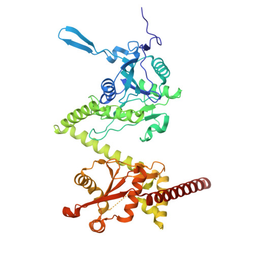
Self-assembling protein cages are versatile nanoscale architectures with broad applications in drug delivery, vaccine development, and structural biology. Historically, two main strategies have been used to construct such cages: genetic fusion of oligomeric domains connected by helical linkers, and computational interface design using either physics-based or machine learning-based methods. Here, we extend the original fusion approach using modern AI algorithms and more sophisticated treatments of helix bending to create protein cages with novel architectures composed exclusively of trimeric building blocks arranged in tetrahedral symmetry. Of 15 designs tested experimentally, multiple sequence variants of two of these designs assembled predominantly into soluble, monodisperse particles of the expected size, with native molecular masses of 633 kDa (T33-Fus-1A, B) and 638 kDa (T33-Fus-2). Cryo-electron microscopy (cryo-EM) structures of three distinct sequence variants spanning from 3.0 to 3.9 Å in resolution confirmed the intended structures in atomic detail, with C-α RMSD values over the entire assemblies as low as 2 Å. The predicted modes of helix bending were similarly validated. The results highlight the impact of methodological improvements for achieving a level of regularity and design precision that has largely evaded prior applications of the fusion approach. These findings expand the prospects and accessible design space for self-assembling protein nanomaterials.
- Structural Biology Programme, Spanish National Cancer Research Centre (CNIO), Madrid28029, Spain.
Organizational Affiliation: