Download Mendeley
Structure and biochemistry reveal substrate-modulated ComEC nuclease activity during DNA processing.
Deselaers, S., Wang, D., Cairoli, T., Afanasyev, P., Hospenthal, M.K.(2026) Nucleic Acids Res 54
- PubMed: 42689412 Search on PubMedSearch on PubMed Central
- DOI: https://doi.org/10.1093/nar/gkag861
- Primary Citation Related Structures:
30BT, 30BU, 32KS - PubMed Abstract:
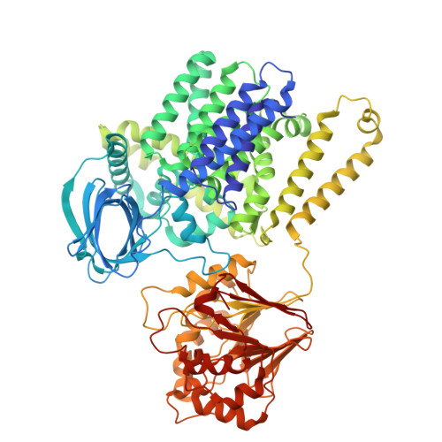
Natural transformation enables bacteria to internalize extracellular DNA, driving adaptation and the spread of antibiotic resistance. The membrane protein ComEC mediates translocation of single-stranded DNA (ssDNA) across the cytoplasmic membrane while degrading the complementary strand, yet the structural basis of its activity remains incompletely defined. Here, we report a cryo-electron microscopy structure of full-length ComEC from Neomoorella carbonis in a pre-translocation state, revealing a three-domain architecture and a conserved transmembrane channel captured in a closed conformation. Structural analysis indicates that conformational rearrangements of channel-lining helices would be required to accommodate ssDNA. Biochemical assays show that, relative to the isolated β-lactamase-like domain, full-length ComEC degrades DNA more efficiently and exhibits position-dependent cleavage of phosphodiester bonds within the DNA substrate. Importantly, coating of the DNA by the periplasmic DNA receptor ComEA suppresses endonucleolytic cleavage and enhances 5'' terminal cleavage, thereby directing ComEC towards productive processing of transforming DNA during natural transformation.
- Institute of Molecular Biology and Biophysics, ETH Zürich, Otto-Stern-Weg 5, 8093 Zürich, Switzerland.
Organizational Affiliation: