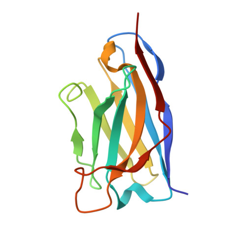
31MR | pdb_000031mr

CryoEM structure of a catalytically inactive CXC Chemokine-degrading protease SpyCEP from Streptococcus pyogenes complexed with an anti-PA-domain monoclonal antibody


Experimental Data Snapshot

  • Method: ELECTRON MICROSCOPY
  • Resolution: 3.07 Å
  • Aggregation State: PARTICLE 
  • Reconstruction Method: SINGLE PARTICLE 

Starting Model: in silico
View more details

wwPDB Validation 3D Report Full Report

Validation slider image for 31MR

This is version 1.1 of the entry. See complete history

Literature

SpyCEP dismantles neutrophil immunity via disorder-driven chemokine remodeling and GAG targeting.

Lau, R.J.Giblin, S.P.Sugar, A.Di Maio, A.Tassini, G.Huse, K.Chorev, D.Chen, Y.Wu, G.H.Huemer, C.B.Kim, S.Y.Matthews, J.Muloud, B.Chen, L.McKenna, S.Xu, Y.Massai, L.Muzzi, C.Ferhati, X.Necchi, F.Gomes Moriel, D.Feizi, T.Liu, Y.Pease, J.E.Sriskandan, S.Matthews, S.

(2026) Proc Natl Acad Sci U S A 123: e2520164123-e2520164123

  • DOI: https://doi.org/10.1073/pnas.2520164123
  • Primary Citation Related Structures: 
    31MR

  • PubMed Abstract: 

    Streptococcus pyogenes (Group A Streptococcus ) employs sophisticated virulence strategies to evade human immunity, including secretion of the cell envelope protease SpyCEP, which cleaves and inactivates key neutrophil-attracting chemokines such as CXCL8. Here, we integrate cryo-electron microscopy, NMR spectroscopy, and native mass spectrometry to investigate how SpyCEP disrupts CXCL8 function. We demonstrate that a disordered aromatic and acidic region within the cleaved autocatalytic maturation loop (CAML) of SpyCEP mimics receptor N-domains and binds an allosteric site on CXCL8. The resulting interaction forms a dynamic fuzzy complex and is coupled to dimer dissociation, consistent with enhanced access to the cleavage site. This disorder-mediated substrate engagement differs from classical protease mechanisms that rely on rigid recognition interfaces. Additionally, glycan microarray and NMR analyses show that the CAML region mediates glycosaminoglycan (GAG) binding, suggesting a means for SpyCEP to maximize encounters with GAG-enriched CXCL8 reservoirs. Together, these findings provide a structural and biophysical framework for understanding how SpyCEP combines substrate engagement with GAG targeting to dismantle chemokine gradients and inhibit neutrophil recruitment. More broadly, this work highlights the role of intrinsic disorder in protease recognition and suggests avenues for anti-virulence therapies and vaccine strategies targeting SpyCEP.


  • Organizational Affiliation
    • Department of Life Sciences, Imperial College London, London SW7 2AZ, United Kingdom.

Macromolecule Content 

  • Total Structure Weight: 198.76 kDa 
  • Atom Count: 12,662 
  • Modeled Residue Count: 1,634 
  • Deposited Residue Count: 1,802 
  • Unique protein chains: 4

Macromolecules

Find similar proteins by:|  3D Structure
Entity ID: 1
MoleculeChains  Sequence LengthOrganismDetailsImage
Cell envelope proteinase A221Streptococcus pyogenesMutation(s): 1 
Gene Names: cepASP119_0326
UniProt
Find proteins for Q201Z7 (Streptococcus pyogenes)
Explore Q201Z7 
Go to UniProtKB:  Q201Z7
Entity Groups
Sequence Clusters30% Identity50% Identity70% Identity90% Identity95% Identity100% Identity
UniProt GroupQ201Z7
Sequence Annotations
Expand
Reference Sequence
Find similar proteins by:|  3D Structure
Entity ID: 2
MoleculeChains  Sequence LengthOrganismDetailsImage
Cell envelope proteinase A1,346Streptococcus pyogenesMutation(s): 2 
Gene Names: cepASP119_0326
UniProt
Find proteins for Q201Z7 (Streptococcus pyogenes)
Explore Q201Z7 
Go to UniProtKB:  Q201Z7
Entity Groups
Sequence Clusters30% Identity50% Identity70% Identity90% Identity95% Identity100% Identity
UniProt GroupQ201Z7
Sequence Annotations
Expand
Reference Sequence
Find similar proteins by:|  3D Structure
Entity ID: 3
MoleculeChains  Sequence LengthOrganismDetailsImage
Anti-PA-domain monoclonal antibody (3F2G10) Heavy chain variable regionC [auth H]122Mus musculusMutation(s): 0 
Entity Groups
Sequence Clusters30% Identity50% Identity70% Identity90% Identity95% Identity100% Identity
Sequence Annotations
Expand
Reference Sequence
Find similar proteins by:|  3D Structure
Entity ID: 4
MoleculeChains  Sequence LengthOrganismDetailsImage
Anti-PA-domain monoclonal antibody (3F2G10) Light chain variable regionD [auth L]113Mus musculusMutation(s): 0 
Entity Groups
Sequence Clusters30% Identity50% Identity70% Identity90% Identity95% Identity100% Identity
Sequence Annotations
Expand
Reference Sequence

Experimental Data & Validation

Experimental Data

  • Method: ELECTRON MICROSCOPY
  • Resolution: 3.07 Å
  • Aggregation State: PARTICLE 
  • Reconstruction Method: SINGLE PARTICLE 
EM Software:
TaskSoftware PackageVersion
RECONSTRUCTIONcryoSPARC
MODEL REFINEMENTISOLDE

Structure Validation

View Full Validation Report


Entry History 

& Funding Information

Deposition Data


Funding OrganizationLocationGrant Number
Medical Research Council (MRC, United Kingdom)United KingdomUKRI682

Revision History  (Full details and data files)

  • Version 1.0: 2026-07-01
    Type: Initial release
  • Version 1.1: 2026-07-22
    Changes: Data collection, Database references