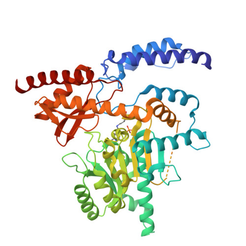
31JF | pdb_000031jf

Human holo aromatic L-amino acid decarboxylase (AADC) D15G variant native structure


Experimental Data Snapshot

  • Method: X-RAY DIFFRACTION
  • Resolution: 2.13 Å
  • R-Value Free: 
    0.217 (Depositor), 0.215 (DCC) 
  • R-Value Work: 
    0.187 (Depositor), 0.186 (DCC) 
  • R-Value Observed: 
    0.189 (Depositor) 

Starting Model: experimental
View more details

wwPDB Validation 3D Report Full Report

Validation slider image for 31JF

This is version 1.0 of the entry. See complete history. 

Literature

Aromatic amino acid decarboxylase - exploring the structural and functional role of its N-terminal domain using deficiency-associated variants.

Bisello, G., Ucar, S.K., Yanbolu, A.Y., Yildirim, C., Perduca, M., D'Ambrosio, M., Franchini, R., Scapin, V., Rampini, M., Bertoldi, M.

(2026) FEBS Lett 

  • DOI: https://doi.org/10.1002/1873-3468.70490
  • Primary Citation Related Structures: 
    31JF

  • PubMed Abstract: 

    AADC deficiency is a rare autosomal recessive disorder characterized by motor/neurodevelopmental symptoms. Approximately one-third of patients experience recurrent hypoglycemia, with or without neurological involvement. To investigate whether specific variants identified in hypoglycemic patients underlie this metabolic phenotype, we characterized three amino-terminal variants. The crystal structure of p.Asp15Gly revealed disruption of an inter-subunit electrostatic network stabilizing the enzyme interface. Computational analyses predicted that p.Val33Leu and p.Gly36Arg perturb an essential catalytic element. Functional studies showed that p.Asp15Gly retained substantial catalytic activity and coenzyme affinity despite reduced stability, whereas p.Val33Leu and p.Gly36Arg exhibited increasing structural destabilization, reduced coenzyme affinity, and severe catalytic defects. These findings reveal distinct functional roles of the amino-terminal-domain, providing a molecular basis for the clinical variability of AADC deficiency.


  • Organizational Affiliation: 
    • Department of Neuroscience, Biomedicine and Movement Sciences, University of Verona, Italy.

Macromolecule Content 

  • Total Structure Weight: 54.52 kDa 
  • Atom Count: 3,673 
  • Modeled Residue Count: 445 
  • Deposited Residue Count: 480 
  • Unique protein chains: 1

Macromolecules

Find similar proteins by:|  3D Structure
Entity ID: 1
MoleculeChains  Sequence LengthOrganismDetailsImage
Aromatic-L-amino-acid decarboxylase480Homo sapiensMutation(s): 2 
Gene Names: DDC, AADC
EC: 4.1.1.28
UniProt & NIH Common Fund Data Resources
Find proteins for P20711 (Homo sapiens)
Explore P20711 
Go to UniProtKB:  P20711
GTEx:  ENSG00000132437 
Entity Groups
Sequence Clusters30% Identity50% Identity70% Identity90% Identity95% Identity100% Identity
UniProt GroupP20711
Sequence Annotations
Expand
Reference Sequence

Small Molecules

Modified Residues  1 Unique
IDChains Modified ResidueModification TypeFormula2D Diagram
LLP
Query on LLP
LYSPyridoxal phosphateC14 H22 N3 O7 P

Experimental Data & Validation

Experimental Data

  • Method: X-RAY DIFFRACTION
  • Resolution: 2.13 Å
  • R-Value Free:  0.217 (Depositor), 0.215 (DCC) 
  • R-Value Work:  0.187 (Depositor), 0.186 (DCC) 
  • R-Value Observed: 0.189 (Depositor) 
Space Group: P 61 2 2
Unit Cell:
Length ( Å )Angle ( ˚ )
a = 106.783α = 90
b = 106.783β = 90
c = 218.377γ = 120
Software Package:
Software NamePurpose
PHENIXrefinement
XDSdata reduction
Aimlessdata scaling
FFTphasing

Structure Validation

View Full Validation Report


Entry History 

& Funding Information

Deposition Data


Funding OrganizationLocationGrant Number
Other privateUnited StatesIIS research grant 2023-2025

Revision History  (Full details and data files)

  • Version 1.0: 2026-11-11
    Type: Initial release