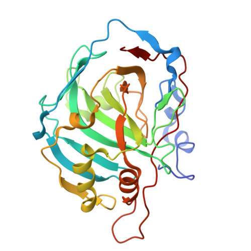
31FP | pdb_000031fp

Crystal structure of carbonic anhydrase I with 4-[2-(ethylsulfanyl)-4-phenyl-1H-imidazol-1-yl]benzene-1-sulfonamide


Experimental Data Snapshot

  • Method: X-RAY DIFFRACTION
  • Resolution: 1.55 Å
  • R-Value Free: 
    0.192 (Depositor), 0.192 (DCC) 
  • R-Value Work: 
    0.162 (Depositor), 0.163 (DCC) 
  • R-Value Observed: 
    0.165 (Depositor) 

Starting Model: experimental
View more details

wwPDB Validation 3D Report Full Report

Validation slider image for 31FP

Ligand Structure Quality Assessment 


This is version 1.0 of the entry. See complete history. 

Literature

Benzenesulfonamide-Imidazole Hybrids as Human Carbonic Anhydrase Inhibitors.

Marcinkeviciute, J., Bagdonas, M., Vainauskas, V., Smirnov, A., Manakova, E., Mickeviciute, A., Juozapaitiene, V., Gedgaudas, M., Mickevicius, V., Matulis, D., Zubriene, A.

(2026) Arch Pharm (Weinheim) 359

  • DOI: https://doi.org/10.1002/ardp.70369
  • Primary Citation Related Structures: 
    31FJ, 31FK, 31FL, 31FM, 31FN, 31FO, 31FP, 31FQ, 31FR, 31FS

  • PubMed Abstract: 

    The high structural similarity among the catalytically active human carbonic anhydrase (CA) isozymes makes the identification of high-affinity, isozyme-selective ligands particularly challenging. This study investigates benzenesulfonamide-imidazole hybrids as inhibitors of CAs and explores structural features of their interactions. Binding affinity was quantified by thermal shift assay, while isothermal titration calorimetry and enzymatic inhibition assays were used to further characterize ligand-CA interactions. Most of the synthesized imidazole-bearing compounds showed high nanomolar affinity for CAII, CAVII, and CAIX isozymes, and their interactions were enthalpically favorable. Crystal structures of CA isozyme-ligand complexes indicate that hydrophobic interactions are the dominant factor in the binding of the examined benzenesulfonamide ligands (4a, 4b, 8, and 12) with CAI, CAII, CAXII, and CAXIII. Combined studies of protein-ligand binding and inhibition, and X-ray crystallography enable analysis of the structural factors responsible for high affinity, which is essential for developing more potent and selective inhibitors for a particular CA isozyme.


  • Organizational Affiliation: 
    • Department of Biothermodynamics and Drug Design, Institute of Biotechnology, Life Sciences Center, Vilnius University, Vilnius, Lithuania.

Macromolecule Content 

  • Total Structure Weight: 59.42 kDa 
  • Atom Count: 4,754 
  • Modeled Residue Count: 513 
  • Deposited Residue Count: 522 
  • Unique protein chains: 1

Macromolecules

Find similar proteins by:|  3D Structure
Entity ID: 1
MoleculeChains  Sequence LengthOrganismDetailsImage
Carbonic anhydrase 1
A, B
261Homo sapiensMutation(s): 0 
Gene Names: CA1
EC: 4.2.1.1 (PDB Primary Data), 4.2.1.69 (UniProt)
UniProt & NIH Common Fund Data Resources
Find proteins for P00915 (Homo sapiens)
Explore P00915 
Go to UniProtKB:  P00915
PHAROS:  P00915
GTEx:  ENSG00000133742 
Entity Groups
Sequence Clusters30% Identity50% Identity70% Identity90% Identity95% Identity100% Identity
UniProt GroupP00915
Sequence Annotations
Expand
Reference Sequence

Small Molecules

Ligands 4 Unique
IDChains Name / Formula / InChI Key2D Diagram3D Interactions
A1J88
(Subject of Investigation/LOI)

Query on A1J88


Download:Ideal Coordinates CCD File
L [auth A],
R [auth B]
4-(2-ethylsulfanyl-4-phenyl-imidazol-1-yl)benzenesulfonamide
C17 H17 N3 O2 S2
DFDNROVYAHGNBV-UHFFFAOYSA-N
DMS

Query on DMS


Download:Ideal Coordinates CCD File
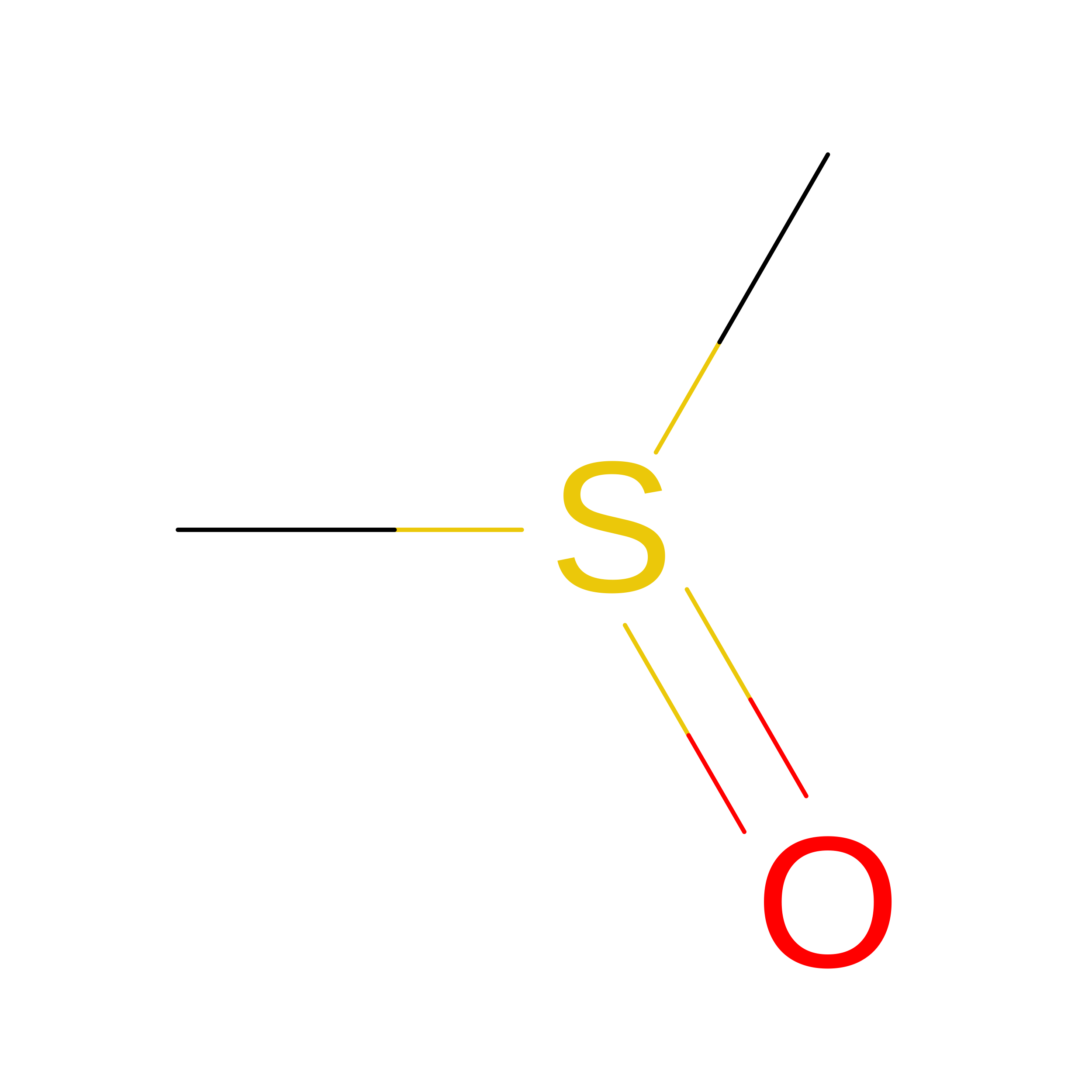
M [auth B]DIMETHYL SULFOXIDE
C2 H6 O S
IAZDPXIOMUYVGZ-UHFFFAOYSA-N
ZN

Query on ZN


Download:Ideal Coordinates CCD File
C [auth A],
N [auth B]
ZINC ION
Zn
PTFCDOFLOPIGGS-UHFFFAOYSA-N
EDO

Query on EDO


Download:Ideal Coordinates CCD File
D [auth A]
E [auth A]
F [auth A]
G [auth A]
H [auth A]
D [auth A],
E [auth A],
F [auth A],
G [auth A],
H [auth A],
I [auth A],
J [auth A],
K [auth A],
O [auth B],
P [auth B],
Q [auth B]
1,2-ETHANEDIOL
C2 H6 O2
LYCAIKOWRPUZTN-UHFFFAOYSA-N

Experimental Data & Validation

Experimental Data

  • Method: X-RAY DIFFRACTION
  • Resolution: 1.55 Å
  • R-Value Free:  0.192 (Depositor), 0.192 (DCC) 
  • R-Value Work:  0.162 (Depositor), 0.163 (DCC) 
  • R-Value Observed: 0.165 (Depositor) 
Space Group: P 21 21 21
Unit Cell:
Length ( Å )Angle ( ˚ )
a = 62.136α = 90
b = 72.993β = 90
c = 121.269γ = 90
Software Package:
Software NamePurpose
REFMACrefinement
Aimlessdata scaling
PDB_EXTRACTdata extraction
XDSdata reduction
MOLREPphasing
Cootmodel building

Structure Validation

View Full Validation Report


Ligand Structure Quality Assessment 


Entry History 

& Funding Information

Deposition Data


Funding OrganizationLocationGrant Number
Not funded--

Revision History  (Full details and data files)

  • Version 1.0: 2026-11-18
    Type: Initial release