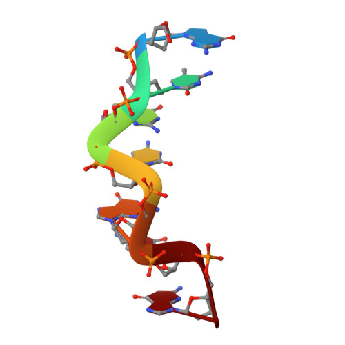
The structures and relative stabilities of d(G x G) reverse Hoogsteen, d(G x T) reverse wobble, and d(G x C) reverse Watson-Crick base-pairs in DNA crystals.
Mooers, B.H., Eichman, B.F., Ho, P.S.(1997) J Mol Biology 269: 796-810
- PubMed: 9223642 Search on PubMed
- DOI: https://doi.org/10.1006/jmbi.1997.1100
- Primary Citation Related Structures:
312D, 313D, 314D - PubMed Abstract:
We have solved the structures of the homoduplex d(Gm5CGCGCG)2, and the heteroduplexes d(GCGCGCG)/d(TCGCGCG) and d(GCGCGCG)/d(CCGCGCG). The structures form six base-pairs of identical Z-DNA duplexes with single nucleotides overhanging at the 5'-ends. The overhanging nucleotide from one strand remains stacked and sandwiched between the blunt-ends of two adjacent Z-DNA duplexes, while the overhanging base of the opposing strand is extra-helical. The stacked and the extra-helical bases from adjacent duplexes pair to form a distorted d(G x G) reverse Hoogsteen base-pair in the d(Gm5CGCGCG)2 homoduplex, and d(G x T) reverse wobble and d(G x C) reverse Watson-Crick base-pairs in the d(GCGCGCG)/d(TCGCGCG) and d(GCGCGCG)/d(CCGCGCG) heteroduplexes, respectively. Interestingly, only the d(G,T) and d(G x C) base-pairs were observed in the heteroduplexes, suggesting that both the d(G x T) reverse wobble and d(G x C) reverse Watson-Crick base-pairs are more stable in this crystal environment than the d(G x G) reverse Hoogsteen base-pair. To estimate the relative stability of the three types of reverse base-pairs, crystals were grown using various mixtures of sequences and their strand compositions analyzed by mass spectrometry. The d(G x C) reverse Watson-Crick base-pair was estimated to be more stable by approximately 1.5 kcal/mol and the d(G x T) reverse wobble base-pair more stable by approximately 0.5 kcal/mol than the d(G x G) reverse Hoogsteen base-pair. The step during crystallization responsible for discriminating between the strands in the crystal is highly cooperative, suggesting that it occurs during the initial nucleating event of crystal growth.
- Department of Biochemistry and Biophysics, Oregon State University, Corvallis 97331, USA.
Organizational Affiliation: