30JK | pdb_000030jk

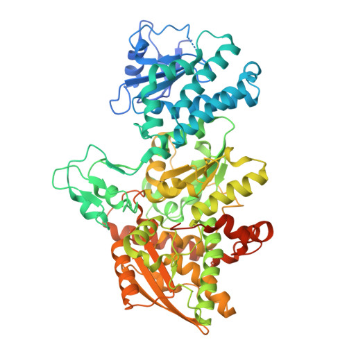
crystal structure of [FeFe]-hydrogenase CbA5H from Clostridium beijerinckii in Hinact state


Experimental Data Snapshot

  • Method: X-RAY DIFFRACTION
  • Resolution: 2.15 Å
  • R-Value Free: 
    0.280 (Depositor), 0.278 (DCC) 
  • R-Value Work: 
    0.249 (Depositor), 0.249 (DCC) 
  • R-Value Observed: 
    0.251 (Depositor) 

Starting Model: experimental
View more details

wwPDB Validation 3D Report Full Report

Validation slider image for 30JK

Ligand Structure Quality Assessment 


This is version 1.1 of the entry. See complete history

Literature

Direct Binding of Cysteine-367 Thiolate to the Active Site of the [FeFe]-Hydrogenase from Clostridium beijerinckii in the O2-Stable State.

Duan, J.Arrigoni, F.Rutz, A.Hofmann, E.Greco, C.Happe, T.

(2026) Biochemistry 65: 2611-2616

  • DOI: https://doi.org/10.1021/acs.biochem.6c00395
  • Primary Citation Related Structures: 
    30JK

  • PubMed Abstract: 

    [FeFe]-hydrogenases are very active biocatalysts for H2 conversion. However, their active site is vulnerable to irreversible degradation initiated by O2 binding at the catalytic iron ion (Fed) of the active center. CbA5H, the [FeFe]-hydrogenase from Clostridium beijerinckii, exhibits stability toward oxygen (O2) due to its ability to reversibly enter an inactive state termed Hinact upon contact with O2. We previously proposed that the close distance of approximately 3.1 Å between the thiol of a nearby cysteine (C367) and Fed, based on a 2.9 Å crystal structure of CbA5H in the Hinact state, enables their binding to each other. This binding therefore was suggested to shield Fed from O2 damage. However, there is currently a lack of evidence to support this hypothesis. Furthermore, density functional theory (DFT) calculations based on a homologous model favored hydroxide as the binding ligand of Fed over the thiol of C367. In this study, we present the crystal structure of CbA5H in the Hinact state at an improved resolution of 2.15 Å. The structure reveals a direct binding between the thiol of C367 and Fed with a distance of approximately 2.77 Å, which is well supported by our DFT calculations based on the new crystallographic data. It is noteworthy that the 2.77 Å bond distance is strikingly long when compared with other iron-sulfur bonds. This finding may provide a crucial foundation for understanding the rapid reversibility of the Hinact state.


  • Organizational Affiliation
    • Photobiotechnology Group, Faculty of Biology and Biotechnology, Ruhr University Bochum, Universitätsstrasse 150, 44801Bochum, Germany.

Macromolecule Content 

  • Total Structure Weight: 155.8 kDa 
  • Atom Count: 10,171 
  • Modeled Residue Count: 1,192 
  • Deposited Residue Count: 1,348 
  • Unique protein chains: 1

Macromolecules

Find similar proteins by:|  3D Structure
Entity ID: 1
MoleculeChains  Sequence LengthOrganismDetailsImage
[FeFe]-hydrogenase
A, B
674Clostridium beijerinckiiMutation(s): 0 
UniProt
Find proteins for A0A1I9RYV3 (Clostridium beijerinckii)
Explore A0A1I9RYV3 
Go to UniProtKB:  A0A1I9RYV3
Entity Groups
Sequence Clusters30% Identity50% Identity70% Identity90% Identity95% Identity100% Identity
UniProt GroupA0A1I9RYV3
Sequence Annotations
Expand
Reference Sequence

Small Molecules

Ligands 6 Unique
IDChains Name / Formula / InChI Key2D Diagram3D Interactions
402
(Subject of Investigation/LOI)

Query on 402


Download:Ideal Coordinates CCD File
C [auth A],
Y [auth B]
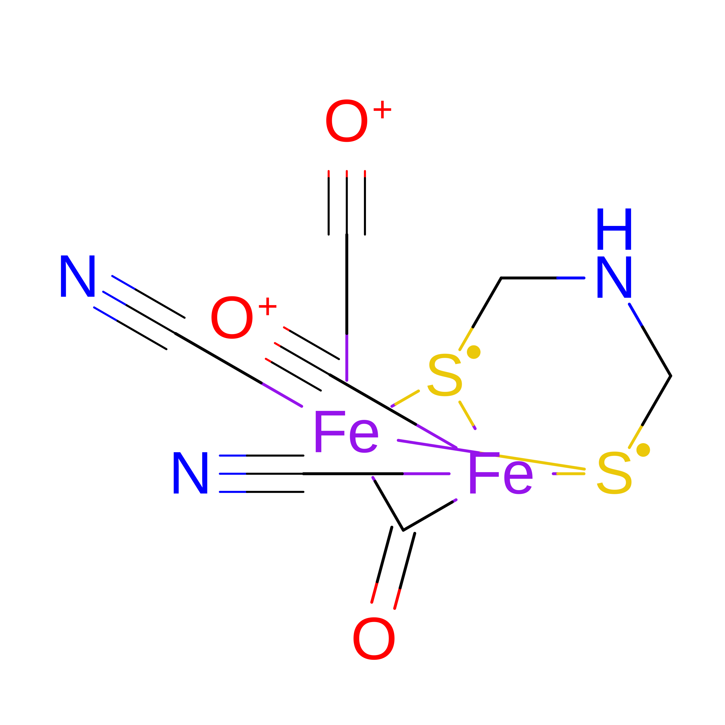
dicarbonyl[bis(cyanide-kappaC)]-mu-(iminodimethanethiolatato-1kappaS:2kappaS)-mu-(oxomethylidene)diiron(2+)
C7 H5 Fe2 N3 O3 S2
LJPDYWPSPOWMIB-UHFFFAOYSA-N
SF4

Query on SF4


Download:Ideal Coordinates CCD File
AA [auth B]
BA [auth B]
D [auth A]
E [auth A]
F [auth A]
AA [auth B],
BA [auth B],
D [auth A],
E [auth A],
F [auth A],
Z [auth B]
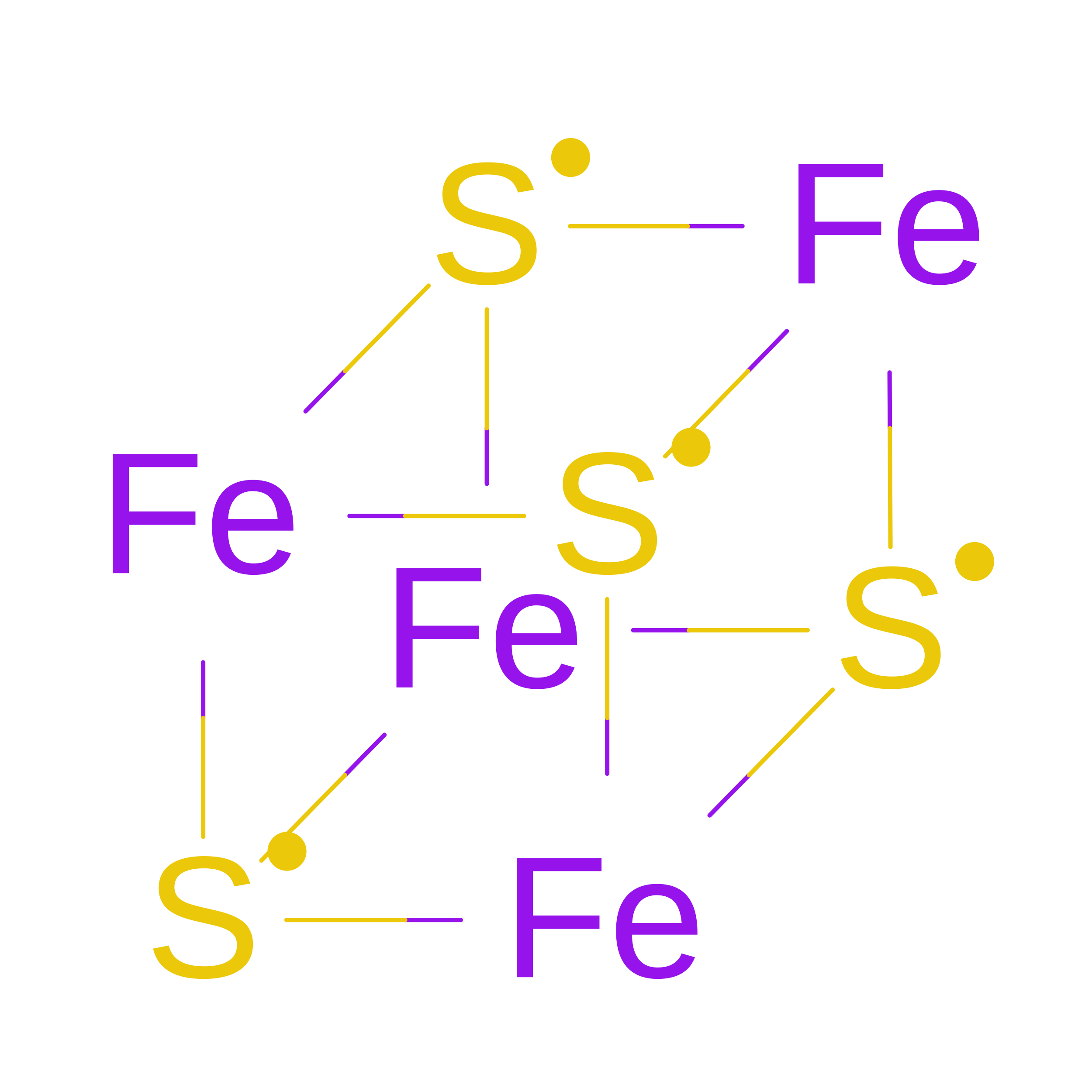
IRON/SULFUR CLUSTER
Fe4 S4
LJBDFODJNLIPKO-UHFFFAOYSA-N
UNK

Query on UNK


Download:Ideal Coordinates CCD File
JA [auth B]
KA [auth B]
LA [auth B]
M [auth A]
MA [auth B]
JA [auth B],
KA [auth B],
LA [auth B],
M [auth A],
MA [auth B],
N [auth A],
NA [auth B],
O [auth A],
OA [auth B],
P [auth A],
PA [auth B],
Q [auth A],
QA [auth B],
R [auth A],
RA [auth B],
S [auth A],
SA [auth B],
T [auth A],
TA [auth B],
U [auth A],
UA [auth B],
V [auth A],
W [auth A],
X [auth A]
UNKNOWN
C4 H9 N O2
QWCKQJZIFLGMSD-VKHMYHEASA-N
ZN

Query on ZN


Download:Ideal Coordinates CCD File
CA [auth B],
G [auth A]
ZINC ION
Zn
PTFCDOFLOPIGGS-UHFFFAOYSA-N
CL

Query on CL


Download:Ideal Coordinates CCD File
EA [auth B]
FA [auth B]
GA [auth B]
HA [auth B]
I [auth A]
EA [auth B],
FA [auth B],
GA [auth B],
HA [auth B],
I [auth A],
IA [auth B],
J [auth A],
K [auth A],
L [auth A]
CHLORIDE ION
Cl
VEXZGXHMUGYJMC-UHFFFAOYSA-M
MG

Query on MG


Download:Ideal Coordinates CCD File
DA [auth B],
H [auth A]
MAGNESIUM ION
Mg
JLVVSXFLKOJNIY-UHFFFAOYSA-N

Experimental Data & Validation

Experimental Data

  • Method: X-RAY DIFFRACTION
  • Resolution: 2.15 Å
  • R-Value Free:  0.280 (Depositor), 0.278 (DCC) 
  • R-Value Work:  0.249 (Depositor), 0.249 (DCC) 
  • R-Value Observed: 0.251 (Depositor) 
Space Group: P 42 21 2
Unit Cell:
Length ( Å )Angle ( ˚ )
a = 168.42α = 90
b = 168.42β = 90
c = 126.31γ = 90
Software Package:
Software NamePurpose
PHENIXrefinement
XSCALEdata scaling
PDB_EXTRACTdata extraction
XDSdata reduction
PHENIXphasing

Structure Validation

View Full Validation Report


Ligand Structure Quality Assessment 


Entry History 

& Funding Information

Deposition Data


Funding OrganizationLocationGrant Number
German Research Foundation (DFG)Germany--

Revision History  (Full details and data files)

  • Version 1.0: 2026-08-12
    Type: Initial release
  • Version 1.1: 2026-08-26
    Changes: Database references