
 30HW | pdb_000030hw

Crystal structure of Apo OrthoTag


Experimental Data Snapshot

  • Method: X-RAY DIFFRACTION
  • Resolution: 1.40 Å
  • R-Value Free: 
    0.185 (Depositor), 0.185 (DCC) 
  • R-Value Work: 
    0.166 (Depositor), 0.166 (DCC) 
  • R-Value Observed: 
    0.167 (Depositor) 

Starting Model: experimental
View more details

wwPDB Validation 3D Report Full Report

Validation slider image for 30HW

This is version 1.0 of the entry. See complete history. 

Literature

Design and Evolution of an Orthogonal HaloTag for Multiplexed Labeling in Cells.

Goldberg, B.J., Lampkin, B.J., Stead, A.T., Stanten, S., Rabe, P., Kritzer, J.A.

(2026) bioRxiv 

  • DOI: https://doi.org/10.64898/2026.05.14.725131
  • Primary Citation Related Structures: 
    30HW

  • PubMed Abstract: 

    The self-labeling protein HaloTag is used to install a wide variety of functional small molecules in cells and living organisms with exquisite specificity with respect to cell type and subcellular localization. HaloTag is a core part of many biotechnology-based tools for sensing, tracking, and manipulating biological systems with a high degree of spatial and temporal control. Due to the limitations of fluorescent proteins and other self-labeling proteins, most of these tools have historically been restricted to a single channel. In this work, we used structure-guided rational design and directed evolution to produce an orthogonal HaloTag protein called OrthoTag which reacts selectively with a modified chloroalkane substrate. OrthoTag retains many of HaloTag's superior properties, and reaction rate measurements with purified proteins show OrthoTag and its substrate have 109-fold mutual orthogonality to HaloTag with respect to substrate concentration. We demonstrate the application of OrthoTag for multiplexed labeling experiments in mammalian cells with minimal optimization. Going forward, OrthoTag should be straightforward to incorporate into HaloTag-based systems to allow simultaneous measurement or manipulation of two biological targets or processes. The availability of multiple high-performance self-labeling proteins will enable the continued development of new multiplexed biotechnology methods.

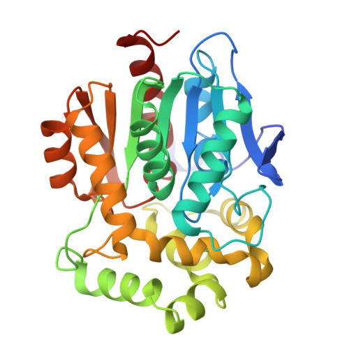
Macromolecule Content 

  • Total Structure Weight: 33.63 kDa 
  • Atom Count: 2,731 
  • Modeled Residue Count: 297 
  • Deposited Residue Count: 297 
  • Unique protein chains: 1

Macromolecules

Find similar proteins by:|  3D Structure
Entity ID: 1
MoleculeChains  Sequence LengthOrganismDetailsImage
Haloalkane dehalogenase297Rhodococcus sp. #1Mutation(s): 24 
Gene Names: dhaA
EC: 3.8.1.5
UniProt
Find proteins for P0A3G3 (Rhodococcus sp)
Explore P0A3G3 
Go to UniProtKB:  P0A3G3
Entity Groups
Sequence Clusters30% Identity50% Identity70% Identity90% Identity95% Identity100% Identity
UniProt GroupP0A3G3
Sequence Annotations
Expand
Reference Sequence

Small Molecules

Ligands 1 Unique
IDChains Name / Formula / InChI Key2D Diagram3D Interactions
ACT

Query on ACT


Download:Ideal Coordinates CCD File
B [auth A]ACETATE ION
C2 H3 O2
QTBSBXVTEAMEQO-UHFFFAOYSA-M

Experimental Data & Validation

Experimental Data

  • Method: X-RAY DIFFRACTION
  • Resolution: 1.40 Å
  • R-Value Free:  0.185 (Depositor), 0.185 (DCC) 
  • R-Value Work:  0.166 (Depositor), 0.166 (DCC) 
  • R-Value Observed: 0.167 (Depositor) 
Space Group: P 1 21 1
Unit Cell:
Length ( Å )Angle ( ˚ )
a = 43.479α = 90
b = 69.918β = 111.68
c = 47.602γ = 90
Software Package:
Software NamePurpose
PHENIXrefinement
PDB_EXTRACTdata extraction
DIALSdata reduction
DIALSdata scaling
PHASERphasing

Structure Validation

View Full Validation Report


Entry History 

& Funding Information

Deposition Data


Funding OrganizationLocationGrant Number
National Institutes of Health/National Institute of General Medical Sciences (NIH/NIGMS)United StatesR35GM148407
Wellcome TrustUnited Kingdom227298/Z/23/Z

Revision History  (Full details and data files)

  • Version 1.0: 2026-10-21
    Type: Initial release