2ZCI | pdb_00002zci

Structure of a GTP-dependent bacterial PEP-carboxykinase from Corynebacterium glutamicum


Experimental Data Snapshot

  • Method: X-RAY DIFFRACTION
  • Resolution: 2.30 Å
  • R-Value Free: 
    0.279 (Depositor), 0.279 (DCC) 
  • R-Value Work: 
    0.188 (Depositor), 0.189 (DCC) 
  • R-Value Observed: 
    0.192 (Depositor) 

Starting Model: experimental
View more details

wwPDB Validation 3D Report Full Report

Validation slider image for 2ZCI

This is version 1.2 of the entry. See complete history

Literature

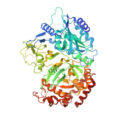
Structure of a GTP-dependent bacterial PEP-carboxykinase from Corynebacterium glutamicum.

Aich, S.Prasad, L.Delbaere, L.T.

(2008) Int J Biochem Cell Biol 40: 1597-1603

  • DOI: https://doi.org/10.1016/j.biocel.2007.12.002
  • Primary Citation Related Structures: 
    2ZCI

  • PubMed Abstract: 

    GTP-dependent phosphoenolpyruvate carboxykinase (PCK) is the key enzyme that controls the blood glucose level during fasting in higher animals. Here we report the first substrate-free structure of a GTP-dependent phosphoenolpyruvate (PEP) carboxykinase from a bacterium, Corynebacterium glutamicum (CgPCK). The protein crystallizes in space group P2(1) with four molecules per asymmetric unit. The 2.3A resolution structure was solved by molecular replacement using the human cytosolic PCK (hcPCK) structure (PDB ID: 1KHF) as the starting model. The four molecules in the asymmetric unit pack as two dimers, and is an artifact of crystal packing. However, the P-loop and the guanine binding loop of the substrate-free CgPCK structure have different conformations from the other published GTP-specific PCK structures, which all have bound substrates and/or metal ions. It appears that a change in the P-loop and guanine binding loop conformation is necessary for substrate binding in GTP-specific PCKs, as opposed to overall domain movement in ATP-specific PCKs.


  • Organizational Affiliation
    • Department of Biochemistry, University of Saskatchewan, Saskatoon, Saskatchewan S7N 5E5, Canada.

Macromolecule Content 

  • Total Structure Weight: 267.75 kDa 
  • Atom Count: 18,407 
  • Modeled Residue Count: 2,334 
  • Deposited Residue Count: 2,440 
  • Unique protein chains: 1

Macromolecules

Find similar proteins by:|  3D Structure
Entity ID: 1
MoleculeChains  Sequence LengthOrganismDetailsImage
Phosphoenolpyruvate carboxykinase [GTP]
A, B, C, D
610Corynebacterium glutamicumMutation(s): 0 
EC: 4.1.1.32
UniProt
Find proteins for Q9AEM1 (Corynebacterium glutamicum (strain ATCC 13032 / DSM 20300 / JCM 1318 / BCRC 11384 / CCUG 27702 / LMG 3730 / NBRC 12168 / NCIMB 10025 / NRRL B-2784 / 534))
Explore Q9AEM1 
Go to UniProtKB:  Q9AEM1
Entity Groups
Sequence Clusters30% Identity50% Identity70% Identity90% Identity95% Identity100% Identity
UniProt GroupQ9AEM1
Sequence Annotations
Expand
Reference Sequence

Experimental Data & Validation

Experimental Data

  • Method: X-RAY DIFFRACTION
  • Resolution: 2.30 Å
  • R-Value Free:  0.279 (Depositor), 0.279 (DCC) 
  • R-Value Work:  0.188 (Depositor), 0.189 (DCC) 
  • R-Value Observed: 0.192 (Depositor) 
Space Group: P 1 21 1
Unit Cell:
Length ( Å )Angle ( ˚ )
a = 70.336α = 90
b = 118.055β = 96.44
c = 152.931γ = 90
Software Package:
Software NamePurpose
ADSCdata collection
REFMACrefinement
HKL-2000data reduction
SCALEPACKdata scaling

Structure Validation

View Full Validation Report


Entry History 

Deposition Data

Revision History  (Full details and data files)

  • Version 1.0: 2008-04-15
    Type: Initial release
  • Version 1.1: 2011-07-13
    Changes: Version format compliance
  • Version 1.2: 2023-08-30
    Changes: Data collection, Database references, Refinement description