2YOR | pdb_00002yor

Crystallization of a 45 kDa peroxygenase- peroxidase from the mushroom Agrocybe aegerita and structure determination by SAD utilizing only the haem iron


Experimental Data Snapshot

  • Method: X-RAY DIFFRACTION
  • Resolution: 2.19 Å
  • R-Value Free: 
    0.237 (Depositor), 0.237 (DCC) 
  • R-Value Work: 
    0.169 (Depositor), 0.170 (DCC) 
  • R-Value Observed: 
    0.172 (Depositor) 

wwPDB Validation 3D Report Full Report

Validation slider image for 2YOR

Ligand Structure Quality Assessment 


This is version 2.1 of the entry. See complete history

Literature

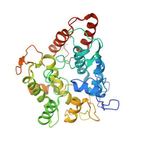
Structural Basis of Substrate Conversion in a New Aromatic Peroxygenase: P450 Functionality with Benefits

Piontek, K.Strittmatter, E.Ullrich, R.Grobe, G.Pecyna, M.J.Kluge, M.Scheibner, K.Hofrichter, M.Plattner, D.A.

(2013) J Biological Chem 288: 34767

  • DOI: https://doi.org/10.1074/jbc.M113.514521
  • Primary Citation Related Structures: 
    2YOR, 2YP1

  • PubMed Abstract: 

    Aromatic peroxygenases (APOs) represent a unique oxidoreductase sub-subclass of heme proteins with peroxygenase and peroxidase activity and were thus recently assigned a distinct EC classification (EC 1.11.2.1). They catalyze, inter alia, oxyfunctionalization reactions of aromatic and aliphatic hydrocarbons with remarkable regio- and stereoselectivities. When compared with cytochrome P450, APOs appear to be the choice enzymes for oxyfunctionalizations in organic synthesis due to their independence from a cellular environment and their greater chemical versatility. Here, the first two crystal structures of a heavily glycosylated fungal aromatic peroxygenase (AaeAPO) are described. They reveal different pH-dependent ligand binding modes. We model the fitting of various substrates in AaeAPO, illustrating the way the enzyme oxygenates polycyclic aromatic hydrocarbons. Spatial restrictions by a phenylalanine pentad in the active-site environment govern substrate specificity in AaeAPO.


  • Organizational Affiliation
    • From the Institute of Organic Chemistry, University of Freiburg, Albertstrasse 21, 79104 Freiburg.

Macromolecule Content 

  • Total Structure Weight: 80.35 kDa 
  • Atom Count: 6,261 
  • Modeled Residue Count: 648 
  • Deposited Residue Count: 650 
  • Unique protein chains: 1

Macromolecules

Find similar proteins by:|  3D Structure
Entity ID: 1
MoleculeChains  Sequence LengthOrganismDetailsImage
AROMATIC PEROXYGENASE
A, B
325Cyclocybe aegeritaMutation(s): 0 
EC: 1.11.2.1
UniProt
Find proteins for B9W4V6 (Cyclocybe aegerita)
Explore B9W4V6 
Go to UniProtKB:  B9W4V6
Entity Groups
Sequence Clusters30% Identity50% Identity70% Identity90% Identity95% Identity100% Identity
UniProt GroupB9W4V6
Glycosylation
Glycosylation Sites: 6
Sequence Annotations
Expand
Reference Sequence

Oligosaccharides

Help  
Entity ID: 2
MoleculeChains Length2D Diagram GlycosylationD Interactions
beta-D-mannopyranose-(1-4)-2-acetamido-2-deoxy-beta-D-glucopyranose-(1-4)-2-acetamido-2-deoxy-beta-D-glucopyranose
C, E
3N-Glycosylation
Glycosylation Resources
GlyTouCan: G15407YE
GlyCosmos: G15407YE
GlyGen: G15407YE
Entity ID: 3
MoleculeChains Length2D Diagram GlycosylationD Interactions
alpha-D-mannopyranose-(1-3)-[alpha-D-mannopyranose-(1-6)]beta-D-mannopyranose-(1-4)-2-acetamido-2-deoxy-beta-D-glucopyranose-(1-4)-2-acetamido-2-deoxy-beta-D-glucopyranose
D
5N-Glycosylation
Glycosylation Resources
GlyTouCan: G22768VO
GlyCosmos: G22768VO
GlyGen: G22768VO
Entity ID: 4
MoleculeChains Length2D Diagram GlycosylationD Interactions
alpha-D-mannopyranose-(1-2)-alpha-D-mannopyranose-(1-6)-[alpha-D-mannopyranose-(1-3)]alpha-D-mannopyranose-(1-6)-[alpha-D-mannopyranose-(1-3)]beta-D-mannopyranose-(1-4)-2-acetamido-2-deoxy-beta-D-glucopyranose-(1-4)-2-acetamido-2-deoxy-beta-D-glucopyranose
F
8N-Glycosylation
Glycosylation Resources
GlyTouCan: G61846BY
GlyCosmos: G61846BY
GlyGen: G61846BY
Entity ID: 5
MoleculeChains Length2D Diagram GlycosylationD Interactions
2-acetamido-2-deoxy-beta-D-glucopyranose-(1-4)-2-acetamido-2-deoxy-beta-D-glucopyranose
G, H, I, J
2N-Glycosylation
Glycosylation Resources
GlyTouCan: G42666HT
GlyCosmos: G42666HT
GlyGen: G42666HT

Small Molecules

Ligands 8 Unique
IDChains Name / Formula / InChI Key2D Diagram3D Interactions
HEM

Query on HEM


Download:Ideal Coordinates CCD File
K [auth A],
V [auth B]
PROTOPORPHYRIN IX CONTAINING FE
C34 H32 Fe N4 O4
KABFMIBPWCXCRK-RGGAHWMASA-L
NAG

Query on NAG


Download:Ideal Coordinates CCD File
AA [auth B],
N [auth A],
O [auth A],
P [auth A],
Y [auth B]
2-acetamido-2-deoxy-beta-D-glucopyranose
C8 H15 N O6
OVRNDRQMDRJTHS-FMDGEEDCSA-N
BMA

Query on BMA


Download:Ideal Coordinates CCD File
Q [auth A],
Z [auth B]
beta-D-mannopyranose
C6 H12 O6
WQZGKKKJIJFFOK-RWOPYEJCSA-N
TRS

Query on TRS


Download:Ideal Coordinates CCD File
BA [auth B]2-AMINO-2-HYDROXYMETHYL-PROPANE-1,3-DIOL
C4 H12 N O3
LENZDBCJOHFCAS-UHFFFAOYSA-O
MZ0

Query on MZ0


Download:Ideal Coordinates CCD File
M [auth A],
X [auth B]
1H-imidazol-5-ylmethanol
C4 H6 N2 O
QDYTUZCWBJRHKK-UHFFFAOYSA-N
SO4

Query on SO4


Download:Ideal Coordinates CCD File
DA [auth B]
FA [auth B]
GA [auth B]
R [auth A]
T [auth A]
DA [auth B],
FA [auth B],
GA [auth B],
R [auth A],
T [auth A],
U [auth A]
SULFATE ION
O4 S
QAOWNCQODCNURD-UHFFFAOYSA-L
CL

Query on CL


Download:Ideal Coordinates CCD File
CA [auth B],
EA [auth B],
HA [auth B],
S [auth A]
CHLORIDE ION
Cl
VEXZGXHMUGYJMC-UHFFFAOYSA-M
MG

Query on MG


Download:Ideal Coordinates CCD File
L [auth A],
W [auth B]
MAGNESIUM ION
Mg
JLVVSXFLKOJNIY-UHFFFAOYSA-N

Experimental Data & Validation

Experimental Data

  • Method: X-RAY DIFFRACTION
  • Resolution: 2.19 Å
  • R-Value Free:  0.237 (Depositor), 0.237 (DCC) 
  • R-Value Work:  0.169 (Depositor), 0.170 (DCC) 
  • R-Value Observed: 0.172 (Depositor) 
Space Group: P 21 21 21
Unit Cell:
Length ( Å )Angle ( ˚ )
a = 77.29α = 90
b = 95.03β = 90
c = 127.2γ = 90
Software Package:
Software NamePurpose
SOLOMONmodel building
XDSdata scaling
SHELXDphasing
SHELXEphasing
autoSHARPphasing
SOLOMONphasing
REFMACrefinement

Structure Validation

View Full Validation Report


Ligand Structure Quality Assessment 


Entry History 

Deposition Data

Revision History  (Full details and data files)

  • Version 1.0: 2013-10-23
    Type: Initial release
  • Version 1.1: 2013-12-11
    Changes: Database references
  • Version 1.2: 2015-04-01
    Changes: Database references
  • Version 2.0: 2020-07-29
    Type: Remediation
    Reason: Carbohydrate remediation
    Changes: Advisory, Atomic model, Data collection, Derived calculations, Other, Structure summary
  • Version 2.1: 2024-11-13
    Changes: Data collection, Database references, Structure summary