2YJ0

X-ray structure of chemically engineered Mycobacterium tuberculosis Dodecin


Experimental Data Snapshot

  • Method: X-RAY DIFFRACTION
  • Resolution: 2.40 Å
  • R-Value Free: 0.275 
  • R-Value Work: 0.217 
  • R-Value Observed: 0.220 

wwPDB Validation   3D Report Full Report


Ligand Structure Quality Assessment 


This is version 1.1 of the entry. See complete history


Literature

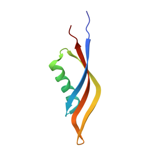
Chemical Engineering of Mycobacterium Tuberculosis Dodecin Hybrids.

Vinzenz, X.Grosse, W.Linne, U.Meissner, B.Essen, L.-O.

(2011) Chem Commun (Camb) 47: 11071

  • DOI: https://doi.org/10.1039/c1cc12929e
  • Primary Citation of Related Structures:  
    2YIZ, 2YJ0

  • PubMed Abstract: 

    The suitability for chemical engineering of the highly symmetrical Mycobacterium tuberculosis dodecin was investigated, its inner cavity providing a large compartment shields introduced compounds from bulk solvent. Hybrids were obtained by S-alkylation of cysteine mutants and characterized by spectroscopic methods, including the crystal structures of wild type and biohybrid dodecins.


  • Organizational Affiliation

    Philipps-Universität Marburg, Fachbereich Chemie, Hans-Meerwein-Strasse, 35032 Marburg, Germany.


Macromolecules
Find similar proteins by:  (by identity cutoff)  |  3D Structure
Entity ID: 1
MoleculeChains Sequence LengthOrganismDetailsImage
DODECIN
A, B, C, D, E
A, B, C, D, E, F
69Mycobacterium tuberculosis H37RvMutation(s): 1 
UniProt
Find proteins for Q8VK10 (Mycobacterium tuberculosis (strain CDC 1551 / Oshkosh))
Explore Q8VK10 
Go to UniProtKB:  Q8VK10
Entity Groups  
Sequence Clusters30% Identity50% Identity70% Identity90% Identity95% Identity100% Identity
UniProt GroupQ8VK10
Sequence Annotations
Expand
  • Reference Sequence
Small Molecules
Ligands 5 Unique
IDChains Name / Formula / InChI Key2D Diagram3D Interactions
COA
Query on COA

Download Ideal Coordinates CCD File 
BA [auth F]
H [auth A]
L [auth B]
Q [auth C]
T [auth D]
BA [auth F],
H [auth A],
L [auth B],
Q [auth C],
T [auth D],
X [auth E]
COENZYME A
C21 H36 N7 O16 P3 S
RGJOEKWQDUBAIZ-IBOSZNHHSA-N
420
Query on 420

Download Ideal Coordinates CCD File 
AA [auth F]
G [auth A]
K [auth B]
P [auth C]
S [auth D]
AA [auth F],
G [auth A],
K [auth B],
P [auth C],
S [auth D],
W [auth E]
N-[2-({[5-(DIMETHYLAMINO)NAPHTHALEN-1-YL]SULFONYL}AMINO)ETHYL]-2-IODOACETAMIDE
C16 H20 I N3 O3 S
QDHGMMDEGVINBU-UHFFFAOYSA-N
FMN
Query on FMN

Download Ideal Coordinates CCD File 
CA [auth F]
I [auth A]
M [auth B]
R [auth C]
U [auth D]
CA [auth F],
I [auth A],
M [auth B],
R [auth C],
U [auth D],
Y [auth E]
FLAVIN MONONUCLEOTIDE
C17 H21 N4 O9 P
FVTCRASFADXXNN-SCRDCRAPSA-N
SO4
Query on SO4

Download Ideal Coordinates CCD File 
V [auth D],
Z [auth E]
SULFATE ION
O4 S
QAOWNCQODCNURD-UHFFFAOYSA-L
CL
Query on CL

Download Ideal Coordinates CCD File 
J [auth A],
N [auth B],
O [auth B]
CHLORIDE ION
Cl
VEXZGXHMUGYJMC-UHFFFAOYSA-M
Experimental Data & Validation

Experimental Data

  • Method: X-RAY DIFFRACTION
  • Resolution: 2.40 Å
  • R-Value Free: 0.275 
  • R-Value Work: 0.217 
  • R-Value Observed: 0.220 
  • Space Group: C 2 2 21
Unit Cell:
Length ( Å )Angle ( ˚ )
a = 94α = 90
b = 103.8β = 90
c = 82.5γ = 90
Software Package:
Software NamePurpose
REFMACrefinement
XDSdata reduction
XSCALEdata scaling
PHASERphasing

Structure Validation

View Full Validation Report


Ligand Structure Quality Assessment 


Entry History 

Deposition Data

Revision History  (Full details and data files)

  • Version 1.0: 2011-10-12
    Type: Initial release
  • Version 1.1: 2023-12-20
    Changes: Data collection, Database references, Derived calculations, Other, Refinement description