2XM3 | pdb_00002xm3

Deinococcus radiodurans ISDra2 Transposase Left end DNA complex


Experimental Data Snapshot

  • Method: X-RAY DIFFRACTION
  • Resolution: 2.30 Å
  • R-Value Free: 
    0.249 (Depositor), 0.242 (DCC) 
  • R-Value Work: 
    0.218 (Depositor), 0.211 (DCC) 
  • R-Value Observed: 
    0.218 (Depositor) 

wwPDB Validation 3D Report Full Report

Validation slider image for 2XM3

This is version 1.4 of the entry. See complete history

Literature

DNA Recognition and the Precleavage State During Single-Stranded DNA Transposition in D. Radiodurans.

Hickman, A.B.James, J.A.Barabas, O.Pasternak, C.Ton-Hoang, B.Chandler, M.Sommer, S.Dyda, F.

(2010) EMBO J 29: 3840

  • DOI: https://doi.org/10.1038/emboj.2010.241
  • Primary Citation Related Structures: 
    2XM3, 2XMA, 2XO6, 2XQC

  • PubMed Abstract: 

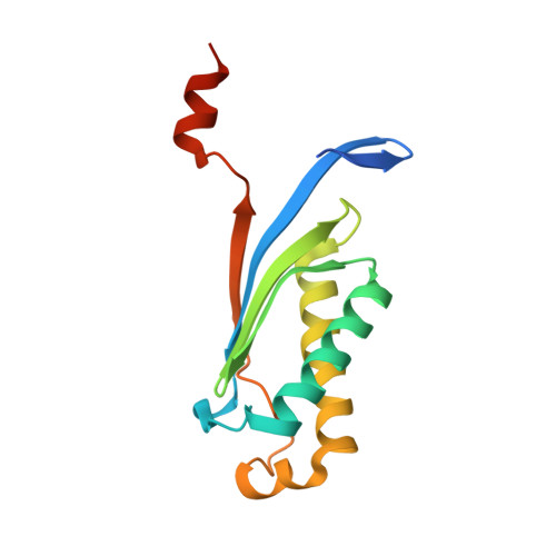
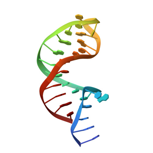
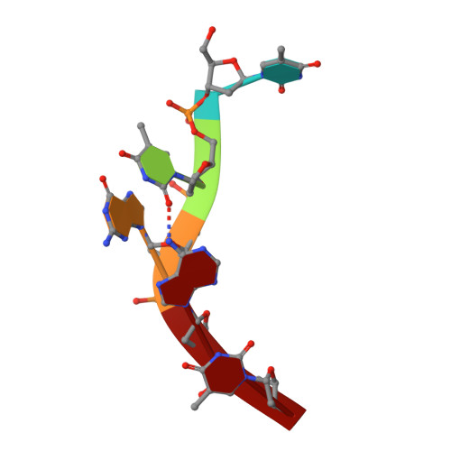
    Bacterial insertion sequences (ISs) from the IS200/IS605 family encode the smallest known DNA transposases and mobilize through single-stranded DNA transposition. Transposition by one particular family member, ISDra2 from Deinococcus radiodurans, is dramatically stimulated upon massive γ irradiation. We have determined the crystal structures of four ISDra2 transposase/IS end complexes; combined with in vivo activity assays and fluorescence anisotropy binding measurements, these have revealed the molecular basis of strand discrimination and transposase action. The structures also show that previously established structural rules of target site recognition that allow different specific sequences to be targeted are only partially conserved among family members. Furthermore, we have captured a fully assembled active site including the scissile phosphate bound by a divalent metal ion cofactor (Cd²(+)) that supports DNA cleavage. Finally, the observed active site rearrangements when the transposase binds a metal ion in which it is inactive provide a clear rationale for metal ion specificity.


  • Organizational Affiliation
    • Laboratory of Molecular Biology, National Institute of Diabetes, Digestive, and Kidney Diseases, National Institutes of Health, Bethesda, MD, USA.

Macromolecule Content 

  • Total Structure Weight: 156.11 kDa 
  • Atom Count: 10,596 
  • Modeled Residue Count: 956 
  • Deposited Residue Count: 1,032 
  • Unique protein chains: 1
  • Unique nucleic acid chains: 2

Macromolecules


Find similar proteins by:|  3D Structure
Entity ID: 1
MoleculeChains  Sequence LengthOrganismDetailsImage
TRANSPOSASE
A, B, C, D, E
A, B, C, D, E, F
140Deinococcus radioduransMutation(s): 0 
UniProt
Find proteins for Q7DF83 (Deinococcus radiodurans (strain ATCC 13939 / DSM 20539 / JCM 16871 / CCUG 27074 / LMG 4051 / NBRC 15346 / NCIMB 9279 / VKM B-1422 / R1))
Explore Q7DF83 
Go to UniProtKB:  Q7DF83
Entity Groups
Sequence Clusters30% Identity50% Identity70% Identity90% Identity95% Identity100% Identity
UniProt GroupQ7DF83
Sequence Annotations
Expand
Reference Sequence
Find similar nucleic acids by:  (by identity cutoff) 
Entity ID: 2
MoleculeChains LengthOrganismImage
DRA2 TRANSPOSASE BINDING ELEMENT
G, I, K, M, O
G, I, K, M, O, Q
27synthetic construct
Sequence Annotations
Expand
Reference Sequence
Find similar nucleic acids by:  Sequence
Entity ID: 3
MoleculeChains LengthOrganismImage
5'-D(*TP*TP*AP*GP*T)-3'
H, J, L, N, P
H, J, L, N, P, R
5synthetic construct
Sequence Annotations
Expand
Reference Sequence

Small Molecules

Ligands 3 Unique
IDChains Name / Formula / InChI Key2D Diagram3D Interactions
GOL

Query on GOL


Download:Ideal Coordinates CCD File
AA [auth J],
DA [auth L],
HA [auth P],
V [auth G]
GLYCEROL
C3 H8 O3
PEDCQBHIVMGVHV-UHFFFAOYSA-N
ACT

Query on ACT


Download:Ideal Coordinates CCD File
S [auth B]ACETATE ION
C2 H3 O2
QTBSBXVTEAMEQO-UHFFFAOYSA-M
MG

Query on MG


Download:Ideal Coordinates CCD File
BA [auth K]
CA [auth K]
EA [auth M]
FA [auth M]
GA [auth O]
BA [auth K],
CA [auth K],
EA [auth M],
FA [auth M],
GA [auth O],
IA [auth Q],
T [auth E],
U [auth F],
W [auth G],
X [auth G],
Y [auth I],
Z [auth I]
MAGNESIUM ION
Mg
JLVVSXFLKOJNIY-UHFFFAOYSA-N

Experimental Data & Validation

Experimental Data

  • Method: X-RAY DIFFRACTION
  • Resolution: 2.30 Å
  • R-Value Free:  0.249 (Depositor), 0.242 (DCC) 
  • R-Value Work:  0.218 (Depositor), 0.211 (DCC) 
  • R-Value Observed: 0.218 (Depositor) 
Space Group: P 21 21 21
Unit Cell:
Length ( Å )Angle ( ˚ )
a = 104.583α = 90
b = 128.384β = 90
c = 140.222γ = 90
Software Package:
Software NamePurpose
CNSrefinement
DENZOdata reduction
SCALEPACKdata scaling
PHASESphasing

Structure Validation

View Full Validation Report


Entry History 

Deposition Data

Revision History  (Full details and data files)

  • Version 1.0: 2010-10-13
    Type: Initial release
  • Version 1.1: 2011-05-08
    Changes: Version format compliance
  • Version 1.2: 2011-07-13
    Changes: Version format compliance
  • Version 1.3: 2019-05-08
    Changes: Data collection, Experimental preparation, Other
  • Version 1.4: 2024-05-08
    Changes: Data collection, Database references, Derived calculations, Other