2X1Q | pdb_00002x1q

Gelsolin Nanobody


Experimental Data Snapshot

  • Method: X-RAY DIFFRACTION
  • Resolution: 1.06 Å
  • R-Value Free: 
    0.229 (Depositor), 0.230 (DCC) 
  • R-Value Work: 
    0.213 (Depositor), 0.214 (DCC) 
  • R-Value Observed: 
    0.214 (Depositor) 

Starting Model: experimental
View more details

wwPDB Validation 3D Report Full Report

Validation slider image for 2X1Q

This is version 1.4 of the entry. See complete history

Literature

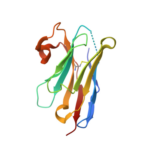
A Llama-Derived Gelsolin Single-Domain Antibody Blocks Gelsolin-G-Actin Interaction.

Van Den Abbeele, A.De Clercq, S.De Ganck, A.De Corte, V.Van Loo, B.Soror, S.H.Srinivasan, V.Steyaert, J.Vandekerckhove, J.Gettemans, J.

(2010) Cell Mol Life Sci 67: 1519

  • DOI: https://doi.org/10.1007/s00018-010-0266-1
  • Primary Citation Related Structures: 
    2X1O, 2X1P, 2X1Q

  • PubMed Abstract: 

    RNA interference has tremendously advanced our understanding of gene function but recent reports have exposed undesirable side-effects. Recombinant Camelid single-domain antibodies (VHHs) provide an attractive means for studying protein function without affecting gene expression. We raised VHHs against gelsolin (GsnVHHs), a multifunctional actin-binding protein that controls cellular actin organization and migration. GsnVHH-induced delocalization of gelsolin to mitochondria or the nucleus in mammalian cells reveals distinct subpopulations including free gelsolin and actin-bound gelsolin complexes. GsnVHH 13 specifically recognizes Ca(2+)-activated gelsolin (K (d) approximately 10 nM) while GsnVHH 11 binds gelsolin irrespective of Ca(2+) (K (d) approximately 5 nM) but completely blocks its interaction with G-actin. Both GsnVHHs trace gelsolin in membrane ruffles of EGF-stimulated MCF-7 cells and delay cell migration without affecting F-actin severing/capping or actin nucleation activities by gelsolin. We conclude that VHHs represent a potent way of blocking structural proteins and that actin nucleation by gelsolin is more complex than previously anticipated.


  • Organizational Affiliation
    • Department of Medical Protein Research, VIB, 9000 Ghent, Belgium.

Macromolecule Content 

  • Total Structure Weight: 27.81 kDa 
  • Atom Count: 2,299 
  • Modeled Residue Count: 240 
  • Deposited Residue Count: 254 
  • Unique protein chains: 1

Macromolecules

Find similar proteins by:|  3D Structure
Entity ID: 1
MoleculeChains  Sequence LengthOrganismDetailsImage
GELSOLIN NANOBODY
A, B
127Lama glamaMutation(s): 0 

Experimental Data & Validation

Experimental Data

  • Method: X-RAY DIFFRACTION
  • Resolution: 1.06 Å
  • R-Value Free:  0.229 (Depositor), 0.230 (DCC) 
  • R-Value Work:  0.213 (Depositor), 0.214 (DCC) 
  • R-Value Observed: 0.214 (Depositor) 
Space Group: P 1 21 1
Unit Cell:
Length ( Å )Angle ( ˚ )
a = 35.908α = 90
b = 63.425β = 90.36
c = 50.01γ = 90
Software Package:
Software NamePurpose
REFMACrefinement
HKLdata reduction
SCALEPACKdata scaling
PHASERphasing

Structure Validation

View Full Validation Report


Entry History 

Revision History  (Full details and data files)

  • Version 1.0: 2011-01-12
    Type: Initial release
  • Version 1.1: 2011-05-08
    Changes: Version format compliance
  • Version 1.2: 2011-07-13
    Changes: Version format compliance
  • Version 1.3: 2023-12-20
    Changes: Data collection, Database references, Other, Refinement description
  • Version 1.4: 2024-11-13
    Changes: Structure summary