2WMM

Crystal structure of the hinge domain of MukB


Experimental Data Snapshot

  • Method: X-RAY DIFFRACTION
  • Resolution: 2.30 Å
  • R-Value Free: 0.240 
  • R-Value Work: 0.217 
  • R-Value Observed: 0.217 

wwPDB Validation   3D Report Full Report


This is version 1.2 of the entry. See complete history


Literature

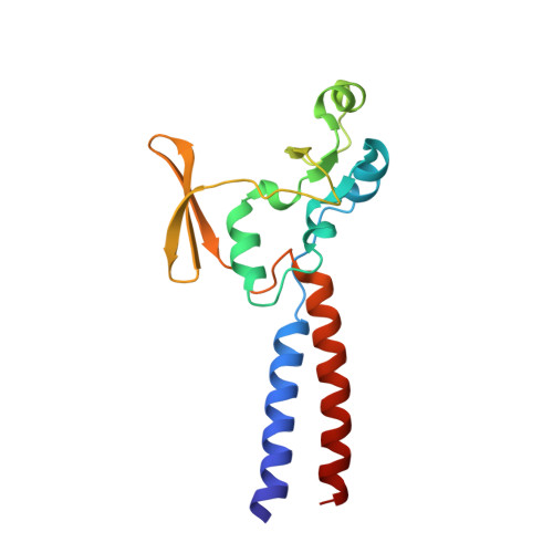
Crystal structure of the MukB hinge domain with coiled-coil stretches and its functional implications.

Ku, B.Lim, J.H.Shin, H.C.Shin, S.Y.Oh, B.H.

(2010) Proteins 78: 1483-1490

  • DOI: https://doi.org/10.1002/prot.22664
  • Primary Citation of Related Structures:  
    2WMM

  • PubMed Abstract: 

    The structural maintenance of chromosomes (SMC) family proteins are commonly found in the multiprotein complexes involved in chromosome organization, including chromosome condensation and sister chromatid cohesion. These proteins are characterized by forming a V-shaped homo- or heterodimeric structure with two long coiled-coil arms having two ATPase head domains at the distal ends. The hinge domain, located in the middle of the coiled coil, forms the dimer interface. In addition to being the dimerization module, SMC hinges appear to play other roles, including the gateway function for DNA entry into the cohesin complex. Herein, we report the homodimeric structure of the hinge domain of Escherichia coli MukB, which forms a prokaryotic condensin complex with two non-SMC subunits, MukE and MukF. In contrast with SMC hinge of Thermotoga maritima which has a sizable central hole at the dimer interface, MukB hinge forms a constricted dimer interface lacking a hole. Under our assay conditions, MukB hinge does not interact with DNA in accordance with the absence of a notable positively charged surface patch. The function of MukB hinge appears to be limited to dimerization of two copies of MukB molecules.


  • Organizational Affiliation

    Department of Biological Sciences, KAIST Institute for the Biocentury, Daejeon, Korea.


Macromolecules
Find similar proteins by:  (by identity cutoff)  |  3D Structure
Entity ID: 1
MoleculeChains Sequence LengthOrganismDetailsImage
Chromosome partition protein MukB
A, B
162Escherichia coli K-12Mutation(s): 1 
Gene Names: mukBb0924JW0907
UniProt
Find proteins for P22523 (Escherichia coli (strain K12))
Explore P22523 
Go to UniProtKB:  P22523
Entity Groups  
Sequence Clusters30% Identity50% Identity70% Identity90% Identity95% Identity100% Identity
UniProt GroupP22523
Sequence Annotations
Expand
  • Reference Sequence
Small Molecules
Ligands 1 Unique
IDChains Name / Formula / InChI Key2D Diagram3D Interactions
MLT
Query on MLT

Download Ideal Coordinates CCD File 
C [auth A]D-MALATE
C4 H6 O5
BJEPYKJPYRNKOW-UWTATZPHSA-N
Experimental Data & Validation

Experimental Data

  • Method: X-RAY DIFFRACTION
  • Resolution: 2.30 Å
  • R-Value Free: 0.240 
  • R-Value Work: 0.217 
  • R-Value Observed: 0.217 
  • Space Group: P 32
Unit Cell:
Length ( Å )Angle ( ˚ )
a = 56.443α = 90
b = 56.443β = 90
c = 115.443γ = 120
Software Package:
Software NamePurpose
CNSrefinement
HKL-2000data reduction
SCALEPACKdata scaling
SOLVEphasing

Structure Validation

View Full Validation Report


Entry History 

Deposition Data

Revision History  (Full details and data files)

  • Version 1.0: 2010-01-12
    Type: Initial release
  • Version 1.1: 2013-01-30
    Changes: Atomic model, Derived calculations, Non-polymer description, Version format compliance
  • Version 1.2: 2018-09-19
    Changes: Data collection, Database references, Source and taxonomy, Structure summary