2WKH | pdb_00002wkh

Crystal structure of the acyl-enzyme OXA-10 K70C-Ampicillin at pH 7


Experimental Data Snapshot

  • Method: X-RAY DIFFRACTION
  • Resolution: 1.79 Å
  • R-Value Free: 
    0.259 (Depositor) 
  • R-Value Work: 
    0.200 (Depositor), 0.202 (DCC) 
  • R-Value Observed: 
    0.203 (Depositor) 

Starting Model: experimental
View more details

wwPDB Validation 3D Report Full Report

Validation slider image for 2WKH

Ligand Structure Quality Assessment 


This is version 1.4 of the entry. See complete history

Literature

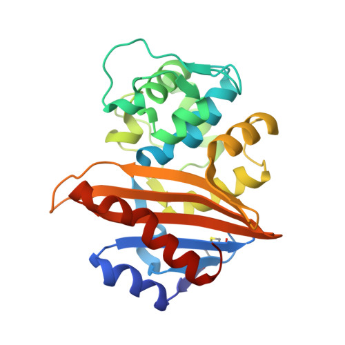
Three Factors that Modulate the Activity of Class D Beta-Lactamases and Interfere with the Post-Translational Carboxylation of Lys70.

Vercheval, L.Bauvois, C.Di Paolo, A.Borel, F.Ferrer, J.Sauvage, E.Matagne, A.Frere, J.Charlier, P.Galleni, M.Kerff, F.

(2010) Biochem J 432: 495

  • DOI: https://doi.org/10.1042/BJ20101122
  • Primary Citation Related Structures: 
    2WGV, 2WGW, 2WKH, 2WKI

  • PubMed Abstract: 

    The activity of class D β-lactamases is dependent on Lys70 carboxylation in the active site. Structural, kinetic and affinity studies show that this post-translational modification can be affected by the presence of a poor substrate such as moxalactam but also by the V117T substitution. Val117 is a strictly conserved hydrophobic residue located in the active site. In addition, inhibition of class D β-lactamases by chloride ions is due to a competition between the side chain carboxylate of the modified Lys70 and chloride ions. Determination of the individual kinetic constants shows that the deacylation of the acyl-enzyme is the rate-limiting step for the wild-type OXA-10 β-lactamase.


  • Organizational Affiliation
    • Laboratory of Biological Macromolecules, Centre for Protein Engineering, Institut de Chimie B6a, University of Liège, Liège 4000, Belgium.

Macromolecule Content 

  • Total Structure Weight: 56.59 kDa 
  • Atom Count: 4,298 
  • Modeled Residue Count: 490 
  • Deposited Residue Count: 496 
  • Unique protein chains: 1

Macromolecules

Find similar proteins by:|  3D Structure
Entity ID: 1
MoleculeChains  Sequence LengthOrganismDetailsImage
BETA-LACTAMASE OXA-10
A, B
248Pseudomonas aeruginosaMutation(s): 1 
EC: 3.5.2.6
UniProt
Find proteins for P14489 (Pseudomonas aeruginosa)
Explore P14489 
Go to UniProtKB:  P14489
Entity Groups
Sequence Clusters30% Identity50% Identity70% Identity90% Identity95% Identity100% Identity
UniProt GroupP14489
Sequence Annotations
Expand
Reference Sequence

Small Molecules

Ligands 2 Unique
IDChains Name / Formula / InChI Key2D Diagram3D Interactions
ZZ7

Query on ZZ7


Download:Ideal Coordinates CCD File
C [auth A],
G [auth B]
(2R,4S)-2-[(R)-{[(2R)-2-amino-2-phenylacetyl]amino}(carboxy)methyl]-5,5-dimethyl-1,3-thiazolidine-4-carboxylic acid
C16 H21 N3 O5 S
KDAWOPKDXRJNHV-MPPDQPJWSA-N
SO4

Query on SO4


Download:Ideal Coordinates CCD File
D [auth A],
E [auth A],
F [auth A],
H [auth B],
I [auth B]
SULFATE ION
O4 S
QAOWNCQODCNURD-UHFFFAOYSA-L

Experimental Data & Validation

Experimental Data

  • Method: X-RAY DIFFRACTION
  • Resolution: 1.79 Å
  • R-Value Free:  0.259 (Depositor) 
  • R-Value Work:  0.200 (Depositor), 0.202 (DCC) 
  • R-Value Observed: 0.203 (Depositor) 
Space Group: P 21 21 21
Unit Cell:
Length ( Å )Angle ( ˚ )
a = 48.92α = 90
b = 97.09β = 90
c = 126.13γ = 90
Software Package:
Software NamePurpose
PHENIXrefinement
XDSdata reduction
SCALAdata scaling
AMoREphasing

Structure Validation

View Full Validation Report


Ligand Structure Quality Assessment 


Entry History 

Deposition Data

Revision History  (Full details and data files)

  • Version 1.0: 2010-08-25
    Type: Initial release
  • Version 1.1: 2012-04-25
    Changes: Database references, Derived calculations, Other, Refinement description, Version format compliance
  • Version 1.2: 2014-10-01
    Changes: Non-polymer description, Structure summary
  • Version 1.3: 2023-12-13
    Changes: Data collection, Database references, Derived calculations, Other, Refinement description
  • Version 1.4: 2024-11-13
    Changes: Structure summary