2VSD | pdb_00002vsd

crystal structure of CHIR-AB1


Experimental Data Snapshot

  • Method: X-RAY DIFFRACTION
  • Resolution: 1.82 Å
  • R-Value Free: 
    0.247 (Depositor), 0.242 (DCC) 
  • R-Value Work: 
    0.207 (Depositor), 0.205 (DCC) 
  • R-Value Observed: 
    0.210 (Depositor) 

Starting Model: experimental
View more details

wwPDB Validation 3D Report Full Report

Validation slider image for 2VSD

This is version 4.2 of the entry. See complete history

Literature

The Crystal Structure of Chir-Ab1: A Primordial Avian Classical Fc Receptor.

Arnon, T.I.Kaiser, J.T.West, A.P.Olson, R.Diskin, R.Viertlboeck, B.C.Gobel, T.W.Bjorkman, P.J.

(2008) J Mol Biology 381: 1012

  • DOI: https://doi.org/10.1016/j.jmb.2008.06.082
  • Primary Citation Related Structures: 
    2VSD

  • PubMed Abstract: 

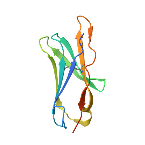
    CHIR-AB1 is a newly identified avian immunoglobulin (Ig) receptor that includes both activating and inhibitory motifs and was therefore classified as a potentially bifunctional receptor. Recently, CHIR-AB1 was shown to bind the Fc region of chicken IgY and to induce calcium mobilization via association with the common gamma-chain, a subunit that transmits signals upon ligation of many different immunoreceptors. Here we describe the 1.8-A-resolution crystal structure of the CHIR-AB1 ectodomain. The receptor ectodomain consists of a single C2-type Ig domain resembling the Ig-like domains found in mammalian Fc receptors such as FcgammaRs and FcalphaRI. Unlike these receptors and other monomeric Ig superfamily members, CHIR-AB1 crystallized as a 2-fold symmetrical homodimer that bears no resemblance to variable or constant region dimers in an antibody. Analytical ultracentrifugation demonstrated that CHIR-AB1 exists as a mixture of monomers and dimers in solution, and equilibrium gel filtration revealed a 2:1 receptor/ligand binding stoichiometry. Measurement of the 1:1 CHIR-AB1/IgY interaction affinity indicates a relatively low affinity complex, but a 2:1 CHIR-AB1/IgY interaction allows an increase in apparent affinity due to avidity effects when the receptor is tethered to a surface. Taken together, these results add to the structural understanding of Fc receptors and their functional mechanisms.


  • Organizational Affiliation
    • Division of Biology, 114-96 and Howard Hughes Medical Institute, California Institute of Technology, Pasadena, CA 91125, USA.

Macromolecule Content 

  • Total Structure Weight: 12.61 kDa 
  • Atom Count: 875 
  • Modeled Residue Count: 93 
  • Deposited Residue Count: 105 
  • Unique protein chains: 1

Macromolecules

Find similar proteins by:|  3D Structure
Entity ID: 1
MoleculeChains  Sequence LengthOrganismDetailsImage
CHIR AB1105Gallus gallusMutation(s): 0 
UniProt
Find proteins for Q5ZJ90 (Gallus gallus)
Explore Q5ZJ90 
Go to UniProtKB:  Q5ZJ90
Entity Groups
Sequence Clusters30% Identity50% Identity70% Identity90% Identity95% Identity100% Identity
UniProt GroupQ5ZJ90
Glycosylation
Glycosylation Sites: 1
Sequence Annotations
Expand
Reference Sequence

Oligosaccharides

Help  
Entity ID: 2
MoleculeChains Length2D Diagram GlycosylationD Interactions
beta-D-mannopyranose-(1-4)-2-acetamido-2-deoxy-beta-D-glucopyranose-(1-4)-2-acetamido-2-deoxy-beta-D-glucopyranose
B
3N-Glycosylation
Glycosylation Resources
GlyTouCan: G15407YE
GlyCosmos: G15407YE
GlyGen: G15407YE

Experimental Data & Validation

Experimental Data

  • Method: X-RAY DIFFRACTION
  • Resolution: 1.82 Å
  • R-Value Free:  0.247 (Depositor), 0.242 (DCC) 
  • R-Value Work:  0.207 (Depositor), 0.205 (DCC) 
  • R-Value Observed: 0.210 (Depositor) 
Space Group: P 31 2 1
Unit Cell:
Length ( Å )Angle ( ˚ )
a = 63.695α = 90
b = 63.695β = 90
c = 55.453γ = 120
Software Package:
Software NamePurpose
PHASERphasing
AMoREphasing
REFMACrefinement

Structure Validation

View Full Validation Report


Entry History 

Deposition Data

Revision History  (Full details and data files)

  • Version 1.0: 2008-07-29
    Type: Initial release
  • Version 1.1: 2011-05-08
    Changes: Version format compliance
  • Version 1.2: 2011-07-13
    Changes: Version format compliance
  • Version 2.0: 2019-09-11
    Changes: Atomic model, Data collection, Derived calculations, Experimental preparation, Non-polymer description, Structure summary
  • Version 3.0: 2019-10-23
    Changes: Advisory, Atomic model, Data collection, Derived calculations, Non-polymer description, Other, Structure summary
  • Version 4.0: 2020-07-29
    Type: Remediation
    Reason: Carbohydrate remediation
    Changes: Advisory, Atomic model, Data collection, Derived calculations, Structure summary
  • Version 4.1: 2023-12-13
    Changes: Data collection, Database references, Derived calculations, Refinement description, Structure summary
  • Version 4.2: 2024-11-20
    Changes: Structure summary