2VGL

AP2 CLATHRIN ADAPTOR CORE


Experimental Data Snapshot

  • Method: X-RAY DIFFRACTION
  • Resolution: 2.60 Å
  • R-Value Free: 0.339 
  • R-Value Work: 0.261 
  • R-Value Observed: 0.265 

wwPDB Validation   3D Report Full Report


Ligand Structure Quality Assessment 


This is version 1.3 of the entry. See complete history


Literature

Molecular Architecture and Functional Model of the Endocytic Ap2 Complex

Collins, B.M.Mccoy, A.J.Kent, H.M.Evans, P.R.Owen, D.J.

(2002) Cell 109: 523

  • DOI: https://doi.org/10.1016/s0092-8674(02)00735-3
  • Primary Citation of Related Structures:  
    2VGL

  • PubMed Abstract: 

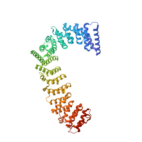
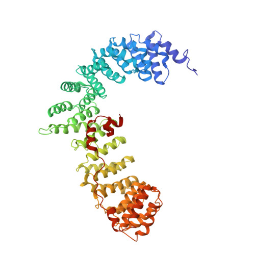
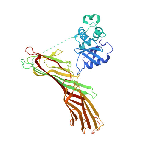
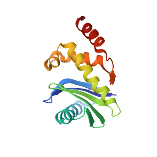
    AP2 is the best-characterized member of the family of heterotetrameric clathrin adaptor complexes that play pivotal roles in many vesicle trafficking pathways within the cell. AP2 functions in clathrin-mediated endocytosis, the process whereby cargo enters the endosomal system from the plasma membrane. We describe the structure of the 200 kDa AP2 "core" (alpha trunk, beta2 trunk, mu2, and sigma2) complexed with the polyphosphatidylinositol headgroup mimic inositolhexakisphosphate at 2.6 A resolution. Two potential polyphosphatidylinositide binding sites are observed, one on alpha and one on mu2. The binding site for Yxxphi endocytic motifs is buried, indicating that a conformational change, probably triggered by phosphorylation in the disordered mu2 linker, is necessary to allow Yxxphi motif binding. A model for AP2 recruitment and activation is proposed.


  • Organizational Affiliation

    Cambridge Institute for Medical Research, University of Cambridge, Department of Clinical Biochemistry, Wellcome Trust/MRC Building, Hills Road, United Kingdom.


Macromolecules
Find similar proteins by:  (by identity cutoff)  |  3D Structure
Entity ID: 1
MoleculeChains Sequence LengthOrganismDetailsImage
ADAPTOR PROTEIN COMPLEX AP-2, ALPHA 2 SUBUNIT621Rattus norvegicusMutation(s): 0 
UniProt
Find proteins for P18484 (Rattus norvegicus)
Explore P18484 
Go to UniProtKB:  P18484
Entity Groups  
Sequence Clusters30% Identity50% Identity70% Identity90% Identity95% Identity100% Identity
UniProt GroupP18484
Sequence Annotations
Expand
  • Reference Sequence
Find similar proteins by:  (by identity cutoff)  |  3D Structure
Entity ID: 2
MoleculeChains Sequence LengthOrganismDetailsImage
AP-2 COMPLEX SUBUNIT BETA-1591Homo sapiensMutation(s): 0 
UniProt & NIH Common Fund Data Resources
Find proteins for P63010 (Homo sapiens)
Explore P63010 
Go to UniProtKB:  P63010
PHAROS:  P63010
GTEx:  ENSG00000006125 
Entity Groups  
Sequence Clusters30% Identity50% Identity70% Identity90% Identity95% Identity100% Identity
UniProt GroupP63010
Sequence Annotations
Expand
  • Reference Sequence
Find similar proteins by:  (by identity cutoff)  |  3D Structure
Entity ID: 3
MoleculeChains Sequence LengthOrganismDetailsImage
AP-2 COMPLEX SUBUNIT MU-1C [auth M]435Rattus norvegicusMutation(s): 0 
UniProt
Find proteins for P84092 (Rattus norvegicus)
Explore P84092 
Go to UniProtKB:  P84092
Entity Groups  
Sequence Clusters30% Identity50% Identity70% Identity90% Identity95% Identity100% Identity
UniProt GroupP84092
Sequence Annotations
Expand
  • Reference Sequence
Find similar proteins by:  (by identity cutoff)  |  3D Structure
Entity ID: 4
MoleculeChains Sequence LengthOrganismDetailsImage
AP-2 COMPLEX SUBUNIT SIGMA-1D [auth S]142Mus musculusMutation(s): 0 
UniProt & NIH Common Fund Data Resources
Find proteins for P62743 (Mus musculus)
Explore P62743 
Go to UniProtKB:  P62743
IMPC:  MGI:2141861
Entity Groups  
Sequence Clusters30% Identity50% Identity70% Identity90% Identity95% Identity100% Identity
UniProt GroupP62743
Sequence Annotations
Expand
  • Reference Sequence
Small Molecules
Ligands 1 Unique
IDChains Name / Formula / InChI Key2D Diagram3D Interactions
IHP
Query on IHP

Download Ideal Coordinates CCD File 
E [auth A]INOSITOL HEXAKISPHOSPHATE
C6 H18 O24 P6
IMQLKJBTEOYOSI-GPIVLXJGSA-N
Experimental Data & Validation

Experimental Data

  • Method: X-RAY DIFFRACTION
  • Resolution: 2.60 Å
  • R-Value Free: 0.339 
  • R-Value Work: 0.261 
  • R-Value Observed: 0.265 
  • Space Group: P 31 2 1
Unit Cell:
Length ( Å )Angle ( ˚ )
a = 121.746α = 90
b = 121.746β = 90
c = 258.124γ = 120
Software Package:
Software NamePurpose
REFMACrefinement
MOSFLMdata reduction
SCALAdata scaling
SHARPphasing

Structure Validation

View Full Validation Report


Ligand Structure Quality Assessment 


Entry History 

Deposition Data

Revision History  (Full details and data files)

  • Version 1.0: 2007-12-25
    Type: Initial release
  • Version 1.1: 2011-05-08
    Changes: Version format compliance
  • Version 1.2: 2011-07-13
    Changes: Version format compliance
  • Version 1.3: 2020-10-07
    Changes: Structure summary