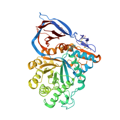
2V3D

acid-beta-glucosidase with N-butyl-deoxynojirimycin


Experimental Data Snapshot

  • Method: X-RAY DIFFRACTION
  • Resolution: 1.96 Å
  • R-Value Free: 0.208 
  • R-Value Work: 0.154 
  • R-Value Observed: 0.157 

Starting Model: experimental
View more details

wwPDB Validation   3D Report Full Report


Ligand Structure Quality Assessment 


This is version 2.1 of the entry. See complete history


Literature

Crystal Structures of Complexes of N-Butyl- and N-Nonyl-Deoxynojirimycin Bound to Acid Beta-Glucosidase: Insights Into the Mechanism of Chemical Chaperone Action in Gaucher Disease.

Brumshtein, B.Greenblatt, H.M.Butters, T.D.Shaaltiel, Y.Aviezer, D.Silman, I.Futerman, A.H.Sussman, J.L.

(2007) J Biol Chem 282: 29052

  • DOI: https://doi.org/10.1074/jbc.M705005200
  • Primary Citation of Related Structures:  
    2V3D, 2V3E

  • PubMed Abstract: 

    Gaucher disease is caused by mutations in the gene encoding acid beta-glucosidase (GlcCerase), resulting in glucosylceramide (GlcCer) accumulation. The only currently available orally administered treatment for Gaucher disease is N-butyl-deoxynojirimycin (Zavesca, NB-DNJ), which partially inhibits GlcCer synthesis, thus reducing levels of GlcCer accumulation. NB-DNJ also acts as a chemical chaperone for GlcCerase, although at a different concentration than that required to completely inhibit GlcCer synthesis. We now report the crystal structures, at 2A resolution, of complexes of NB-DNJ and N-nonyl-deoxynojirimycin (NN-DNJ) with recombinant human GlcCerase, expressed in cultured plant cells. Both inhibitors bind at the active site of GlcCerase, with the imino sugar moiety making hydrogen bonds to side chains of active site residues. The alkyl chains of NB-DNJ and NN-DNJ are oriented toward the entrance of the active site where they undergo hydrophobic interactions. Based on these structures, we make a number of predictions concerning (i) involvement of loops adjacent to the active site in the catalytic process, (ii) the nature of nucleophilic attack by Glu-340, and (iii) the role of a conserved water molecule located in a solvent cavity adjacent to the active site. Together, these results have significance for understanding the mechanism of action of GlcCerase and the mode of GlcCerase chaperoning by imino sugars.


  • Organizational Affiliation

    Departments of Structural Biology, Weizmann Institute of Science, Rehovot 76100, Israel.


Macromolecules
Find similar proteins by:  (by identity cutoff)  |  3D Structure
Entity ID: 1
MoleculeChains Sequence LengthOrganismDetailsImage
GLUCOSYLCERAMIDASE
A, B
505Homo sapiensMutation(s): 0 
EC: 3.2.1.45 (PDB Primary Data), 3.2.1.46 (UniProt), 2.4.1 (UniProt), 3.2.1 (UniProt)
UniProt & NIH Common Fund Data Resources
Find proteins for P04062 (Homo sapiens)
Explore P04062 
Go to UniProtKB:  P04062
PHAROS:  P04062
GTEx:  ENSG00000177628 
Entity Groups  
Sequence Clusters30% Identity50% Identity70% Identity90% Identity95% Identity100% Identity
UniProt GroupP04062
Glycosylation
Glycosylation Sites: 1Go to GlyGen: P04062-1
Sequence Annotations
Expand
  • Reference Sequence
Oligosaccharides

Help

Entity ID: 2
MoleculeChains Length2D Diagram Glycosylation3D Interactions
beta-D-mannopyranose-(1-4)-2-acetamido-2-deoxy-beta-D-glucopyranose-(1-4)-[alpha-L-fucopyranose-(1-3)]2-acetamido-2-deoxy-beta-D-glucopyranose
C, D
4N-Glycosylation
Glycosylation Resources
GlyTouCan:  G18638YB
GlyCosmos:  G18638YB
GlyGen:  G18638YB
Small Molecules
Ligands 3 Unique
IDChains Name / Formula / InChI Key2D Diagram3D Interactions
NAG
Query on NAG

Download Ideal Coordinates CCD File 
K [auth B]2-acetamido-2-deoxy-beta-D-glucopyranose
C8 H15 N O6
OVRNDRQMDRJTHS-FMDGEEDCSA-N
NBV
Query on NBV

Download Ideal Coordinates CCD File 
F [auth A],
L [auth B]
(2R,3R,4R,5S)-1-BUTYL-2-(HYDROXYMETHYL)PIPERIDINE-3,4,5-TRIOL
C10 H21 N O4
UQRORFVVSGFNRO-UTINFBMNSA-N
SO4
Query on SO4

Download Ideal Coordinates CCD File 
E [auth A]
G [auth A]
H [auth A]
I [auth A]
J [auth B]
E [auth A],
G [auth A],
H [auth A],
I [auth A],
J [auth B],
M [auth B],
N [auth B],
O [auth B]
SULFATE ION
O4 S
QAOWNCQODCNURD-UHFFFAOYSA-L
Binding Affinity Annotations 
IDSourceBinding Affinity
NBV PDBBind:  2V3D Ki: 1.16e+5 (nM) from 1 assay(s)
BindingDB:  2V3D Ki: 1.16e+5 (nM) from 1 assay(s)
IC50: min: 1.18e+5, max: 4.00e+5 (nM) from 7 assay(s)
Experimental Data & Validation

Experimental Data

  • Method: X-RAY DIFFRACTION
  • Resolution: 1.96 Å
  • R-Value Free: 0.208 
  • R-Value Work: 0.154 
  • R-Value Observed: 0.157 
  • Space Group: P 1 21 1
Unit Cell:
Length ( Å )Angle ( ˚ )
a = 67.993α = 90
b = 97.21β = 102.91
c = 82.406γ = 90
Software Package:
Software NamePurpose
REFMACrefinement
DENZOdata reduction
SCALEPACKdata scaling
PHASERphasing

Structure Validation

View Full Validation Report


Ligand Structure Quality Assessment 


Entry History 

Deposition Data

Revision History  (Full details and data files)

  • Version 1.0: 2007-08-14
    Type: Initial release
  • Version 1.1: 2011-05-08
    Changes: Version format compliance
  • Version 1.2: 2011-07-13
    Changes: Version format compliance
  • Version 2.0: 2020-07-29
    Type: Remediation
    Reason: Carbohydrate remediation
    Changes: Advisory, Atomic model, Data collection, Derived calculations, Other, Structure summary
  • Version 2.1: 2023-12-13
    Changes: Data collection, Database references, Refinement description, Structure summary