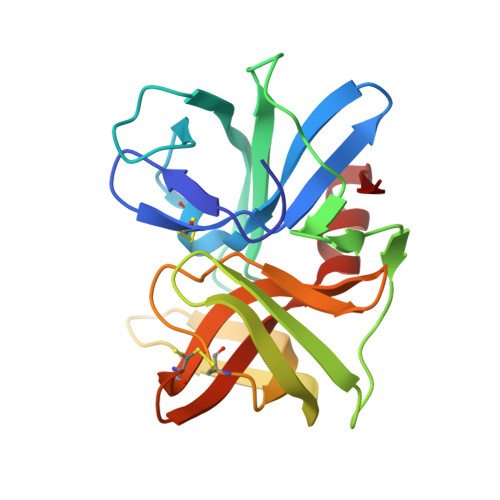
1.2A-resolution crystal structures reveal the second tetrahedral intermediates of streptogrisin B (SGPB).
Lee, T.W., James, M.N.(2008) Biochim Biophys Acta 1784: 319-334
- PubMed: 18157955
- DOI: https://doi.org/10.1016/j.bbapap.2007.11.012
- Primary Citation of Related Structures:
2QA9, 2QAA - PubMed Abstract:
Streptogrisin B (SGPB) has served as one of the models for studying the catalytic activities of serine peptidases. Here we report its native crystal structures at pH 4.2 at a resolution of 1.18A, and at pH 7.3 at a resolution of 1.23A. Unexpectedly, outstanding electron density peaks occurred in the active site and the substrate-binding region of SGPB in the computed maps at both pHs. The densities at pH 4.2 were assigned as a tetrapeptide, Asp-Ala-Ile-Tyr, whereas those at pH 7.3 were assigned as a tyrosine molecule and a leucine molecule existing at equal occupancies in both of the SGPB molecules in the asymmetric unit. Refinement with relaxed geometric restraints resulted in molecular structures representing mixtures of the second tetrahedral intermediates and the enzyme-product complexes of SGPB existing in a pH-dependent equilibrium. Structural comparisons with the complexes of SGPB with turkey ovomucoid third domain (OMTKY3) and its variants have shown that, upon the formation of the tetrahedral intermediate, residues Glu192A to Gly193 of SGPB move towards the alpha-carboxylate O of residue P1 of the bound species, and adjustments in the side-chain conformational angles of His57 and Ser195 of SGPB favor the progression of the catalytic mechanism of SGPB.
- Group in Protein Structure and Function, Department of Biochemistry, University of Alberta, Room 4-29, Medical Sciences Building, Edmonton, Alberta T6G 2H7, Canada.
Organizational Affiliation:


