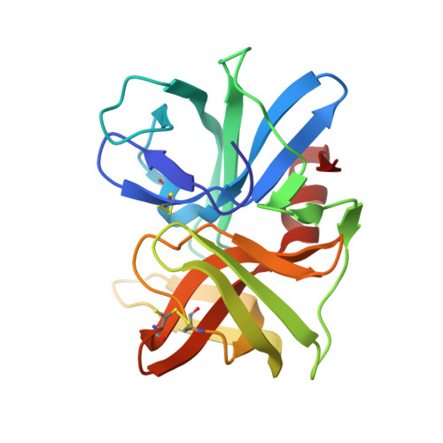
2QAA

Crystal structure of the second tetrahedral intermediates of SGPB at pH 7.3


Experimental Data Snapshot

  • Method: X-RAY DIFFRACTION
  • Resolution: 1.23 Å
  • R-Value Free: 0.127 
  • R-Value Work: 0.113 
  • R-Value Observed: 0.113 

wwPDB Validation   3D Report Full Report


Ligand Structure Quality Assessment 


This is version 1.2 of the entry. See complete history


Literature

1.2A-resolution crystal structures reveal the second tetrahedral intermediates of streptogrisin B (SGPB).

Lee, T.W.James, M.N.

(2008) Biochim Biophys Acta 1784: 319-334

  • DOI: https://doi.org/10.1016/j.bbapap.2007.11.012
  • Primary Citation of Related Structures:  
    2QA9, 2QAA

  • PubMed Abstract: 

    Streptogrisin B (SGPB) has served as one of the models for studying the catalytic activities of serine peptidases. Here we report its native crystal structures at pH 4.2 at a resolution of 1.18A, and at pH 7.3 at a resolution of 1.23A. Unexpectedly, outstanding electron density peaks occurred in the active site and the substrate-binding region of SGPB in the computed maps at both pHs. The densities at pH 4.2 were assigned as a tetrapeptide, Asp-Ala-Ile-Tyr, whereas those at pH 7.3 were assigned as a tyrosine molecule and a leucine molecule existing at equal occupancies in both of the SGPB molecules in the asymmetric unit. Refinement with relaxed geometric restraints resulted in molecular structures representing mixtures of the second tetrahedral intermediates and the enzyme-product complexes of SGPB existing in a pH-dependent equilibrium. Structural comparisons with the complexes of SGPB with turkey ovomucoid third domain (OMTKY3) and its variants have shown that, upon the formation of the tetrahedral intermediate, residues Glu192A to Gly193 of SGPB move towards the alpha-carboxylate O of residue P1 of the bound species, and adjustments in the side-chain conformational angles of His57 and Ser195 of SGPB favor the progression of the catalytic mechanism of SGPB.


  • Organizational Affiliation

    Group in Protein Structure and Function, Department of Biochemistry, University of Alberta, Room 4-29, Medical Sciences Building, Edmonton, Alberta T6G 2H7, Canada.


Macromolecules
Find similar proteins by:  (by identity cutoff)  |  3D Structure
Entity ID: 1
MoleculeChains Sequence LengthOrganismDetailsImage
Streptogrisin-B
A, B
185Streptomyces griseusMutation(s): 0 
EC: 3.4.21.81
UniProt
Find proteins for P00777 (Streptomyces griseus)
Explore P00777 
Go to UniProtKB:  P00777
Entity Groups  
Sequence Clusters30% Identity50% Identity70% Identity90% Identity95% Identity100% Identity
UniProt GroupP00777
Sequence Annotations
Expand
  • Reference Sequence
Small Molecules
Ligands 8 Unique
IDChains Name / Formula / InChI Key2D Diagram3D Interactions
EPE
Query on EPE

Download Ideal Coordinates CCD File 
N [auth A]4-(2-HYDROXYETHYL)-1-PIPERAZINE ETHANESULFONIC ACID
C8 H18 N2 O4 S
JKMHFZQWWAIEOD-UHFFFAOYSA-N
TYR
Query on TYR

Download Ideal Coordinates CCD File 
AA [auth B],
I [auth A]
TYROSINE
C9 H11 N O3
OUYCCCASQSFEME-QMMMGPOBSA-N
LEU
Query on LEU

Download Ideal Coordinates CCD File 
BA [auth B],
J [auth A]
LEUCINE
C6 H13 N O2
ROHFNLRQFUQHCH-YFKPBYRVSA-N
SO4
Query on SO4

Download Ideal Coordinates CCD File 
C [auth A],
D [auth A],
R [auth B],
S [auth B],
T [auth B]
SULFATE ION
O4 S
QAOWNCQODCNURD-UHFFFAOYSA-L
GOL
Query on GOL

Download Ideal Coordinates CCD File 
DA [auth B],
EA [auth B],
FA [auth B],
O [auth A]
GLYCEROL
C3 H8 O3
PEDCQBHIVMGVHV-UHFFFAOYSA-N
EDO
Query on EDO

Download Ideal Coordinates CCD File 
CA [auth B],
K [auth A],
L [auth A],
M [auth A]
1,2-ETHANEDIOL
C2 H6 O2
LYCAIKOWRPUZTN-UHFFFAOYSA-N
ACY
Query on ACY

Download Ideal Coordinates CCD File 
GA [auth B],
P [auth A],
Q [auth A]
ACETIC ACID
C2 H4 O2
QTBSBXVTEAMEQO-UHFFFAOYSA-N
CL
Query on CL

Download Ideal Coordinates CCD File 
E [auth A]
F [auth A]
G [auth A]
H [auth A]
U [auth B]
E [auth A],
F [auth A],
G [auth A],
H [auth A],
U [auth B],
V [auth B],
W [auth B],
X [auth B],
Y [auth B],
Z [auth B]
CHLORIDE ION
Cl
VEXZGXHMUGYJMC-UHFFFAOYSA-M
Experimental Data & Validation

Experimental Data

  • Method: X-RAY DIFFRACTION
  • Resolution: 1.23 Å
  • R-Value Free: 0.127 
  • R-Value Work: 0.113 
  • R-Value Observed: 0.113 
  • Space Group: I 2 3
Unit Cell:
Length ( Å )Angle ( ˚ )
a = 142.286α = 90
b = 142.286β = 90
c = 142.286γ = 90
Software Package:
Software NamePurpose
REFMACrefinement
DENZOdata reduction
SCALEPACKdata scaling
MOLREPphasing

Structure Validation

View Full Validation Report


Ligand Structure Quality Assessment 


Entry History 

Deposition Data

Revision History  (Full details and data files)

  • Version 1.0: 2007-12-11
    Type: Initial release
  • Version 1.1: 2011-07-13
    Changes: Derived calculations, Version format compliance
  • Version 1.2: 2024-11-20
    Changes: Data collection, Database references, Derived calculations, Structure summary