2Q3L | pdb_00002q3l

CRYSTAL STRUCTURE OF AN UNCHARACTERIZED PROTEIN FROM DUF3478 FAMILY WITH A SPOIIAA-LIKE FOLD (SHEW_3102) FROM SHEWANELLA LOIHICA PV-4 AT 2.25 A RESOLUTION


Experimental Data Snapshot

  • Method: X-RAY DIFFRACTION
  • Resolution: 2.25 Å
  • R-Value Free: 
    0.239 (Depositor), 0.235 (DCC) 
  • R-Value Work: 
    0.181 (Depositor), 0.188 (DCC) 
  • R-Value Observed: 
    0.184 (Depositor) 

wwPDB Validation 3D Report Full Report

Validation slider image for 2Q3L

This is version 1.7 of the entry. See complete history

Literature

Open and closed conformations of two SpoIIAA-like proteins (YP_749275.1 and YP_001095227.1) provide insights into membrane association and ligand binding.

Kumar, A.Lomize, A.Jin, K.K.Carlton, D.Miller, M.D.Jaroszewski, L.Abdubek, P.Astakhova, T.Axelrod, H.L.Chiu, H.J.Clayton, T.Das, D.Deller, M.C.Duan, L.Feuerhelm, J.Grant, J.C.Grzechnik, A.Han, G.W.Klock, H.E.Knuth, M.W.Kozbial, P.Krishna, S.S.Marciano, D.McMullan, D.Morse, A.T.Nigoghossian, E.Okach, L.Reyes, R.Rife, C.L.Sefcovic, N.Tien, H.J.Trame, C.B.van den Bedem, H.Weekes, D.Xu, Q.Hodgson, K.O.Wooley, J.Elsliger, M.A.Deacon, A.M.Godzik, A.Lesley, S.A.Wilson, I.A.

(2010) Acta Crystallogr Sect F Struct Biol Cryst Commun 66: 1245-1253

  • DOI: https://doi.org/10.1107/S1744309109042481
  • Primary Citation Related Structures: 
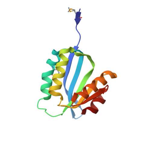
    2OOK, 2Q3L

  • PubMed Abstract: 

    The crystal structures of the proteins encoded by the YP_749275.1 and YP_001095227.1 genes from Shewanella frigidimarina and S. loihica, respectively, have been determined at 1.8 and 2.25 Å resolution, respectively. These proteins are members of a novel family of bacterial proteins that adopt the α/β SpoIIAA-like fold found in STAS and CRAL-TRIO domains. Despite sharing 54% sequence identity, these two proteins adopt distinct conformations arising from different dispositions of their α2 and α3 helices. In the `open' conformation (YP_001095227.1), these helices are 15 Å apart, leading to the creation of a deep nonpolar cavity. In the `closed' structure (YP_749275.1), the helices partially unfold and rearrange, occluding the cavity and decreasing the solvent-exposed hydrophobic surface. These two complementary structures are reminiscent of the conformational switch in CRAL-TRIO carriers of hydrophobic compounds. It is suggested that both proteins may associate with the lipid bilayer in their `open' monomeric state by inserting their amphiphilic helices, α2 and α3, into the lipid bilayer. These bacterial proteins may function as carriers of nonpolar substances or as interfacially activated enzymes.


  • Organizational Affiliation
    • Stanford Synchrotron Radiation Lightsource, SLAC National Accelerator Laboratory, Menlo Park, CA, USA.

Macromolecule Content 

  • Total Structure Weight: 29.73 kDa 
  • Atom Count: 2,133 
  • Modeled Residue Count: 248 
  • Deposited Residue Count: 252 
  • Unique protein chains: 1

Macromolecules

Find similar proteins by:|  3D Structure
Entity ID: 1
MoleculeChains  Sequence LengthOrganismDetailsImage
Uncharacterized protein
A, B
126Shewanella loihica PV-4Mutation(s): 2 
Gene Names: YP_001095227.1Shew_3102
UniProt
Find proteins for A3QHM0 (Shewanella loihica (strain ATCC BAA-1088 / PV-4))
Explore A3QHM0 
Go to UniProtKB:  A3QHM0
Entity Groups
Sequence Clusters30% Identity50% Identity70% Identity90% Identity95% Identity100% Identity
UniProt GroupA3QHM0
Sequence Annotations
Expand
Reference Sequence

Small Molecules

Ligands 3 Unique
IDChains Name / Formula / InChI Key2D Diagram3D Interactions
MPD

Query on MPD


Download:Ideal Coordinates CCD File
E [auth A]
F [auth A]
G [auth A]
H [auth A]
K [auth B]
E [auth A],
F [auth A],
G [auth A],
H [auth A],
K [auth B],
L [auth B],
M [auth B],
N [auth B],
O [auth B]
(4S)-2-METHYL-2,4-PENTANEDIOL
C6 H14 O2
SVTBMSDMJJWYQN-YFKPBYRVSA-N
CL

Query on CL


Download:Ideal Coordinates CCD File
D [auth A],
J [auth B]
CHLORIDE ION
Cl
VEXZGXHMUGYJMC-UHFFFAOYSA-M
NA

Query on NA


Download:Ideal Coordinates CCD File
C [auth A],
I [auth B]
SODIUM ION
Na
FKNQFGJONOIPTF-UHFFFAOYSA-N
Modified Residues  1 Unique
IDChains TypeFormula2D DiagramParent
MSE
Query on MSE
A, B
L-PEPTIDE LINKINGC5 H11 N O2 SeMET

Experimental Data & Validation

Experimental Data

  • Method: X-RAY DIFFRACTION
  • Resolution: 2.25 Å
  • R-Value Free:  0.239 (Depositor), 0.235 (DCC) 
  • R-Value Work:  0.181 (Depositor), 0.188 (DCC) 
  • R-Value Observed: 0.184 (Depositor) 
Space Group: C 2 2 21
Unit Cell:
Length ( Å )Angle ( ˚ )
a = 40.387α = 90
b = 113.963β = 90
c = 130.692γ = 90
Software Package:
Software NamePurpose
REFMACrefinement
PHENIXrefinement
SHELXphasing
MolProbitymodel building
SCALAdata scaling
PDB_EXTRACTdata extraction
MAR345data collection
MOSFLMdata reduction
SHELXDphasing
autoSHARPphasing

Structure Validation

View Full Validation Report


Entry History 

Deposition Data

Revision History  (Full details and data files)

  • Version 1.0: 2007-06-12
    Type: Initial release
  • Version 1.1: 2008-05-01
    Changes: Version format compliance
  • Version 1.2: 2011-07-13
    Changes: Advisory, Version format compliance
  • Version 1.3: 2017-10-18
    Changes: Refinement description
  • Version 1.4: 2017-10-25
    Changes: Author supporting evidence
  • Version 1.5: 2019-07-24
    Changes: Data collection, Derived calculations, Refinement description
  • Version 1.6: 2023-01-25
    Changes: Database references, Derived calculations
  • Version 1.7: 2024-11-20
    Changes: Data collection, Refinement description, Structure summary