2NUL | pdb_00002nul

PEPTIDYLPROLYL ISOMERASE FROM E. COLI


Experimental Data Snapshot

  • Method: X-RAY DIFFRACTION
  • Resolution: 2.10 Å
  • R-Value Work: 
    0.160 (Depositor), 0.157 (DCC) 
  • R-Value Observed: 
    0.160 (Depositor) 

Starting Model: experimental
View more details

wwPDB Validation 3D Report Full Report

Validation slider image for 2NUL

This is version 2.1 of the entry. See complete history

Literature

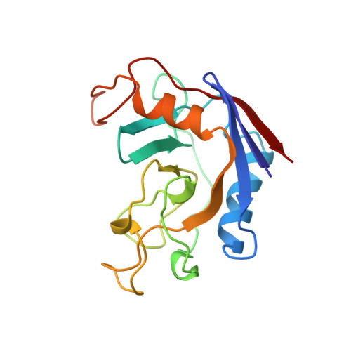
Crystal structure of cytoplasmic Escherichia coli peptidyl-prolyl isomerase: evidence for decreased mobility of loops upon complexation.

Edwards, K.J.Ollis, D.L.Dixon, N.E.

(1997) J Mol Biology 271: 258-265

  • DOI: https://doi.org/10.1006/jmbi.1997.1151
  • Primary Citation Related Structures: 
    2NUL

  • PubMed Abstract: 

    The structure of the unliganded form of the Escherichia coli cytoplasmic peptidyl-prolyl isomerase (ppiB gene product) in a new crystal form was determined by the molecular replacement method and refined to an R-factor of 16.1% at 2.1 A resolution. The enzyme crystallized in the orthorhombic C2221 space group with unit cell dimensions of a=44.7 A, b=68.2 A and c=102.0 A. Comparison with the reported structure of the enzyme complexed with the tripeptide substrate succinyl-Ala-Pro-Ala-p-nitroanilide revealed subtle changes that occur upon complex formation. There is evidence to suggest that two surface loops have significantly reduced mobility in the complexed structure.


  • Organizational Affiliation
    • Research School of Chemistry, Australian National University, Canberra, ACT 0200, Australia.

Macromolecule Content 

  • Total Structure Weight: 18.17 kDa 
  • Atom Count: 1,306 
  • Modeled Residue Count: 163 
  • Deposited Residue Count: 164 
  • Unique protein chains: 1

Macromolecules

Find similar proteins by:|  3D Structure
Entity ID: 1
MoleculeChains  Sequence LengthOrganismDetailsImage
PEPTIDYLPROLYL ISOMERASE164Escherichia coliMutation(s): 0 
EC: 5.2.1.8
UniProt
Find proteins for P23869 (Escherichia coli (strain K12))
Explore P23869 
Go to UniProtKB:  P23869
Entity Groups
Sequence Clusters30% Identity50% Identity70% Identity90% Identity95% Identity100% Identity
UniProt GroupP23869
Sequence Annotations
Expand
Reference Sequence

Experimental Data & Validation

Experimental Data

  • Method: X-RAY DIFFRACTION
  • Resolution: 2.10 Å
  • R-Value Work:  0.160 (Depositor), 0.157 (DCC) 
  • R-Value Observed: 0.160 (Depositor) 
Space Group: C 2 2 21
Unit Cell:
Length ( Å )Angle ( ˚ )
a = 44.7α = 90
b = 68.2β = 90
c = 102γ = 90
Software Package:
Software NamePurpose
X-PLORmodel building
X-PLORrefinement
RIGAKUdata reduction
RIGAKUdata scaling
X-PLORphasing

Structure Validation

View Full Validation Report


Entry History 

Deposition Data

Revision History  (Full details and data files)

  • Version 1.0: 1997-11-19
    Type: Initial release
  • Version 1.1: 2008-03-24
    Changes: Version format compliance
  • Version 1.2: 2011-07-13
    Changes: Version format compliance
  • Version 1.3: 2011-11-16
    Changes: Atomic model
  • Version 2.0: 2023-08-09
    Changes: Atomic model, Database references, Derived calculations, Other, Refinement description
  • Version 2.1: 2024-05-22
    Changes: Data collection