Download Mendeley
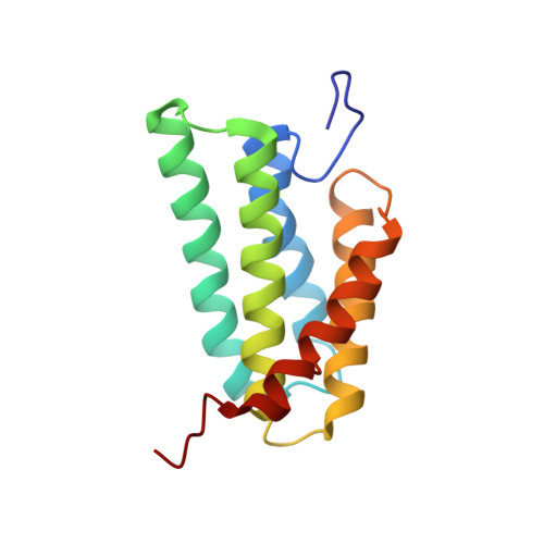
Sequential pH-driven dimerization and stabilization of the N-terminal domain enables rapid spider silk formation.
Kronqvist, N., Otikovs, M., Chmyrov, V., Chen, G., Andersson, M., Nordling, K., Landreh, M., Sarr, M., Jornvall, H., Wennmalm, S., Widengren, J., Meng, Q., Rising, A., Otzen, D., Knight, S.D., Jaudzems, K., Johansson, J.(2014) Nat Commun 5: 3254-3254
- PubMed: 24510122 Search on PubMed
- DOI: https://doi.org/10.1038/ncomms4254
- Primary Citation Related Structures:
2LTH - PubMed Abstract:
The mechanisms controlling the conversion of spider silk proteins into insoluble fibres, which happens in a fraction of a second and in a defined region of the silk glands, are still unresolved. The N-terminal domain changes conformation and forms a homodimer when pH is lowered from 7 to 6; however, the molecular details still remain to be determined. Here we investigate site-directed mutants of the N-terminal domain from Euprosthenops australis major ampullate spidroin 1 and find that the charged residues D40, R60 and K65 mediate intersubunit electrostatic interactions. Protonation of E79 and E119 is required for structural conversions of the subunits into a dimer conformation, and subsequent protonation of E84 around pH 5.7 leads to the formation of a fully stable dimer. These residues are highly conserved, indicating that the now proposed three-step mechanism prevents premature aggregation of spidroins and enables fast formation of spider silk fibres in general.
- KI Alzheimer Disease Research Centre, NVS Department, Karolinska Institutet, Novum, 5th floor, SE-141 86 Stockholm, Sweden.
Organizational Affiliation: