2LMC | pdb_00002lmc

Structure of T7 transcription factor Gp2-E. coli RNAp jaw domain complex


Experimental Data Snapshot

  • Method: SOLUTION NMR
  • Conformers Calculated: 100 
  • Conformers Submitted: 10 
  • Selection Criteria: structures with the lowest energy 

wwPDB Validation 3D Report Full Report

Validation slider image for 2LMC

This is version 1.3 of the entry. See complete history

Literature

Structural and Mechanistic Basis for the Inhibition of Escherichia coli RNA Polymerase by T7 Gp2.

James, E.Liu, M.Sheppard, C.Mekler, V.Camara, B.Liu, B.Simpson, P.Cota, E.Severinov, K.Matthews, S.Wigneshweraraj, S.

(2012) Mol Cell 47: 755-766

  • DOI: https://doi.org/10.1016/j.molcel.2012.06.013
  • Primary Citation Related Structures: 
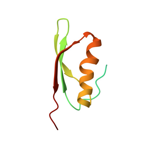
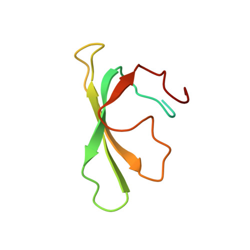
    2LMC

  • PubMed Abstract: 

    The T7 phage-encoded small protein Gp2 is a non-DNA-binding transcription factor that interacts with the jaw domain of the Escherichia coli (Ec) RNA polymerase (RNAp) β' subunit and inhibits transcriptionally proficient promoter-complex (RPo) formation. Here, we describe the high-resolution solution structure of the Gp2-Ec β' jaw domain complex and show that Gp2 and DNA compete for binding to the β' jaw domain. We reveal that efficient inhibition of RPo formation by Gp2 requires the amino-terminal σ(70) domain region 1.1 (R1.1), and that Gp2 antagonizes the obligatory movement of R1.1 during RPo formation. We demonstrate that Gp2 inhibits RPo formation not just by steric occlusion of the RNAp-DNA interaction but also through long-range antagonistic effects on RNAp-promoter interactions around the RNAp active center that likely occur due to repositioning of R1.1 by Gp2. The inhibition of Ec RNAp by Gp2 thus defines a previously uncharacterized mechanism by which bacterial transcription is regulated by a viral factor.


  • Organizational Affiliation
    • MRC Centre for Molecular Bacteriology and Infection, Imperial College London, London SW7 2AZ, UK.

Macromolecule Content 

  • Total Structure Weight: 18.71 kDa 
  • Atom Count: 947 
  • Modeled Residue Count: 120 
  • Deposited Residue Count: 168 
  • Unique protein chains: 2

Macromolecules

Find similar proteins by:|  3D Structure
Entity ID: 1
MoleculeChains  Sequence LengthOrganismDetailsImage
Bacterial RNA polymerase inhibitor84Escherichia phage T7Mutation(s): 0 
Gene Names: 2Gp2
UniProt
Find proteins for P03704 (Escherichia phage T7)
Explore P03704 
Go to UniProtKB:  P03704
Entity Groups
Sequence Clusters30% Identity50% Identity70% Identity90% Identity95% Identity100% Identity
UniProt GroupP03704
Sequence Annotations
Expand
Reference Sequence
Find similar proteins by:|  3D Structure
Entity ID: 2
MoleculeChains  Sequence LengthOrganismDetailsImage
DNA-directed RNA polymerase subunit beta84Escherichia coli K-12Mutation(s): 0 
Gene Names: b3988JW3951RNAPrpoCtabB
EC: 2.7.7.6
UniProt
Find proteins for P0A8T7 (Escherichia coli (strain K12))
Explore P0A8T7 
Go to UniProtKB:  P0A8T7
Entity Groups
Sequence Clusters30% Identity50% Identity70% Identity90% Identity95% Identity100% Identity
UniProt GroupP0A8T7
Sequence Annotations
Expand
Reference Sequence

Experimental Data & Validation

Experimental Data

  • Method: SOLUTION NMR
  • Conformers Calculated: 100 
  • Conformers Submitted: 10 
  • Selection Criteria: structures with the lowest energy 

Structure Validation

View Full Validation Report


Entry History 

Deposition Data

  • Released Date: 2012-03-14 
  • Deposition Author(s): Liu, M.

Revision History  (Full details and data files)

  • Version 1.0: 2012-03-14
    Type: Initial release
  • Version 1.1: 2012-08-01
    Changes: Database references
  • Version 1.2: 2012-10-03
    Changes: Database references
  • Version 1.3: 2024-05-01
    Changes: Data collection, Database references