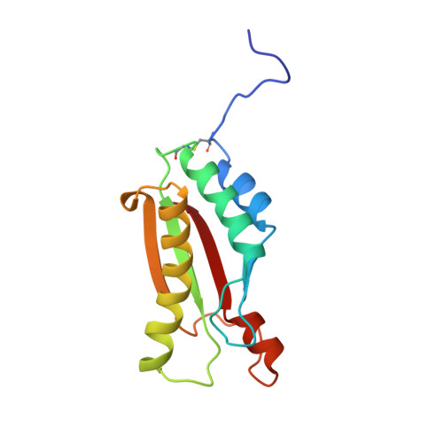
Structure of the Mycobacterium tuberculosis OmpATb protein: A model of an oligomeric channel in the mycobacterial cell wall.
Yang, Y., Auguin, D., Delbecq, S., Dumas, E., Molle, G., Molle, V., Roumestand, C., Saint, N.(2011) Proteins 79: 645-661
- PubMed: 21117233
- DOI: https://doi.org/10.1002/prot.22912
- Primary Citation of Related Structures:
2KGS, 2KGW - PubMed Abstract:
The pore-forming outer membrane protein OmpATb from Mycobacterium tuberculosis is a virulence factor required for acid resistance in host phagosomes. In this study, we determined the 3D structure of OmpATb by NMR in solution. We found that OmpATb is composed of two independent domains separated by a proline-rich hinge region. As expected, the high-resolution structure of the C-terminal domain (OmpATb(198-326)) revealed a module structurally related to other OmpA-like proteins from Gram-negative bacteria. The N-terminal domain of OmpATb (73-204), which is sufficient to form channels in planar lipid bilayers, exhibits a fold, which belongs to the α+β sandwich class fold. Its peculiarity is to be composed of two overlapping subdomains linked via a BON (Bacterial OsmY and Nodulation) domain initially identified in bacterial proteins predicted to interact with phospholipids. Although OmpATb(73-204) is highly water soluble, current-voltage measurements demonstrate that it is able to form conducting pores in model membranes. A HADDOCK modeling of the NMR data gathered on the major monomeric form and on the minor oligomeric populations of OmpATb(73-204) suggest that OmpATb(73-204) can form oligomeric rings able to insert into phospholipid membrane, similar to related proteins from the Type III secretion systems, which form multisubunits membrane-associated rings at the basal body of the secretion machinery.
- Centre de Biochimie Structurale, CNRS UMR 5048, Université Montpellier 1 et 2, F34090 Montpellier, France.
Organizational Affiliation:


