2JBX | pdb_00002jbx

Crystal Structure of the myxoma virus anti-apoptotic protein M11L


Experimental Data Snapshot

  • Method: X-RAY DIFFRACTION
  • Resolution: 2.73 Å
  • R-Value Free: 
    0.286 (Depositor), 0.235 (DCC) 
  • R-Value Work: 
    0.214 (Depositor), 0.218 (DCC) 
  • R-Value Observed: 
    0.218 (Depositor) 

Starting Model: other
View more details

wwPDB Validation 3D Report Full Report

Validation slider image for 2JBX

This is version 1.4 of the entry. See complete history

Literature

A Structural Viral Mimic of Prosurvival Bcl-2: A Pivotal Role for Sequestering Proapoptotic Bax and Bak.

Kvansakul, M.Van Delft, M.F.Lee, E.F.Gulbis, J.M.Fairlie, W.D.Huang, D.C.S.Colman, P.M.

(2007) Mol Cell 25: 933

  • DOI: https://doi.org/10.1016/j.molcel.2007.02.004
  • Primary Citation Related Structures: 
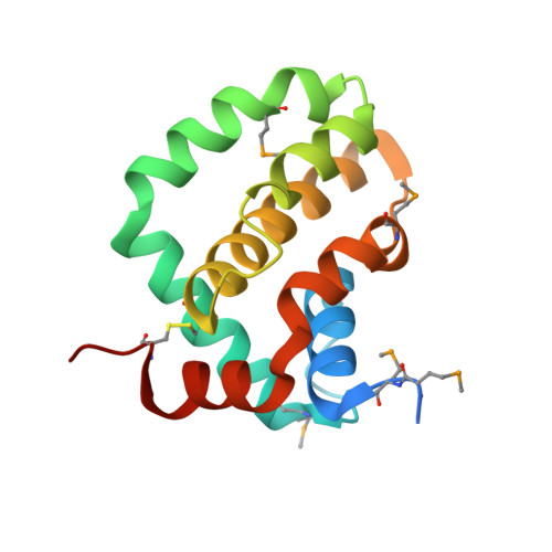
    2JBX, 2JBY

  • PubMed Abstract: 

    Many viruses express antiapoptotic proteins to counter host defense mechanisms that would otherwise trigger the rapid clearance of infected cells. For example, adenoviruses and some gamma-herpesviruses express homologs of prosurvival Bcl-2 to subvert the host's apoptotic machinery. Myxoma virus, a double-stranded DNA virus of the pox family, harbors antiapoptotic M11L, its virulence factor. Analysis of its three-dimensional structure reveals that despite lacking any primary sequence similarity to Bcl-2, it adopts a virtually identical protein fold. This allows it to associate with BH3 domains, especially those of Bax and Bak. We found that M11L acts primarily by sequestering Bax and Bak, thereby blocking the killing action of these essential cell-death mediators. These findings expand the family of protein sequences that act like Bcl-2 to block apoptosis and support the conclusion that the prosurvival action of these proteins critically depends on their ability to bind and antagonize Bax and/or Bak.


  • Organizational Affiliation
    • The Walter and Eliza Hall Institute of Medical Research, 1G Royal Parade, Parkville, Victoria 3050, Australia.

Macromolecule Content 

  • Total Structure Weight: 33.76 kDa 
  • Atom Count: 2,132 
  • Modeled Residue Count: 261 
  • Deposited Residue Count: 290 
  • Unique protein chains: 1

Macromolecules

Find similar proteins by:|  3D Structure
Entity ID: 1
MoleculeChains  Sequence LengthOrganismDetailsImage
M11L PROTEIN
A, B
145Myxoma virusMutation(s): 0 
UniProt
Find proteins for Q85295 (Myxoma virus)
Explore Q85295 
Go to UniProtKB:  Q85295
Entity Groups
Sequence Clusters30% Identity50% Identity70% Identity90% Identity95% Identity100% Identity
UniProt GroupQ85295
Sequence Annotations
Expand
Reference Sequence

Small Molecules

Modified Residues  1 Unique
IDChains TypeFormula2D DiagramParent
MSE
Query on MSE
A, B
L-PEPTIDE LINKINGC5 H11 N O2 SeMET

Experimental Data & Validation

Experimental Data

  • Method: X-RAY DIFFRACTION
  • Resolution: 2.73 Å
  • R-Value Free:  0.286 (Depositor), 0.235 (DCC) 
  • R-Value Work:  0.214 (Depositor), 0.218 (DCC) 
  • R-Value Observed: 0.218 (Depositor) 
Space Group: P 1
Unit Cell:
Length ( Å )Angle ( ˚ )
a = 39.876α = 71.79
b = 39.398β = 90.09
c = 49.222γ = 81.66
Software Package:
Software NamePurpose
REFMACrefinement
HKL-2000data reduction
HKL-2000data scaling
PHASERphasing

Structure Validation

View Full Validation Report


Entry History 

Deposition Data

Revision History  (Full details and data files)

  • Version 1.0: 2007-03-27
    Type: Initial release
  • Version 1.1: 2011-05-08
    Changes: Version format compliance
  • Version 1.2: 2011-07-13
    Changes: Version format compliance
  • Version 1.3: 2024-05-01
    Changes: Data collection, Database references, Derived calculations, Other, Refinement description
  • Version 1.4: 2024-11-20
    Changes: Structure summary