2IIE | pdb_00002iie

single chain Integration Host Factor protein (scIHF2) in complex with DNA


Experimental Data Snapshot

  • Method: X-RAY DIFFRACTION
  • Resolution: 2.41 Å
  • R-Value Free: 
    0.272 (Depositor), 0.276 (DCC) 
  • R-Value Work: 
    0.233 (Depositor), 0.233 (DCC) 
  • R-Value Observed: 
    0.234 (Depositor) 

Starting Model: experimental
View more details

wwPDB Validation 3D Report Full Report

Validation slider image for 2IIE

This is version 1.5 of the entry. See complete history

Literature

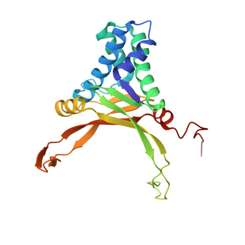
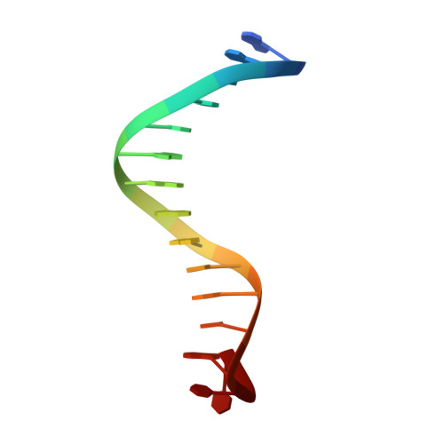
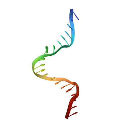
A Divalent Metal-mediated Switch Controlling Protein-induced DNA Bending

Bao, Q.Chen, H.Liu, Y.Yan, J.Droge, P.Davey, C.A.

(2007) J Mol Biology 367: 731-740

  • DOI: https://doi.org/10.1016/j.jmb.2006.09.082
  • Primary Citation Related Structures: 
    2IIE, 2IIF

  • PubMed Abstract: 

    Architectural proteins that reconfigure the paths of DNA segments are required for the establishment of functional interfaces in many genomic transactions. A single-chain derivative of the DNA architectural protein integration host factor was found to adopt two stable conformational states in complex with a specific DNA target. In the so-called open state, the degree of protein-induced DNA bending is reduced significantly compared with the closed state. The conformational switch between these states is controlled by divalent metal binding in two electronegative zones arising from the lysine-to-glutamate substitution in the protein body proximal to the phosphate backbone of one DNA arm. We show that this switch can be employed to control the efficiency of site-specific recombination catalyzed by lambda integrase. Introduction of acidic residues at the protein-DNA interface holds potential for the design of metal-mediated switches for the investigation of functional relationships.


  • Organizational Affiliation
    • Division of Genomics and Genetics, School of Biological Sciences, Nanyang Technological University, Singapore 637551, Singapore.

Macromolecule Content 

  • Total Structure Weight: 44.76 kDa 
  • Atom Count: 3,318 
  • Modeled Residue Count: 274 
  • Deposited Residue Count: 274 
  • Unique protein chains: 1
  • Unique nucleic acid chains: 3

Macromolecules


Find similar proteins by:|  3D Structure
Entity ID: 4
MoleculeChains  Sequence LengthOrganismDetailsImage
Integration host factorD [auth A]204Escherichia coliMutation(s): 0 
UniProt
Find proteins for P0A6X7 (Escherichia coli (strain K12))
Explore P0A6X7 
Go to UniProtKB:  P0A6X7
Find proteins for P0A6Y1 (Escherichia coli (strain K12))
Explore P0A6Y1 
Go to UniProtKB:  P0A6Y1
Entity Groups
Sequence Clusters30% Identity50% Identity70% Identity90% Identity95% Identity100% Identity
UniProt GroupsP0A6X7P0A6Y1
Sequence Annotations
Expand
Reference Sequence
Find similar nucleic acids by:  (by identity cutoff) 
Entity ID: 1
MoleculeChains LengthOrganismImage
Phage P H' siteA [auth C]35synthetic construct
Sequence Annotations
Expand
Reference Sequence
Find similar nucleic acids by:  Sequence
Entity ID: 2
MoleculeChains LengthOrganismImage
DNA (5'-D(*DGP*DGP*DCP*DCP*DAP*DAP*DAP*DAP*DAP*DAP*DGP*DCP*DAP*DTP*DT)-3')B [auth D]15synthetic construct
Sequence Annotations
Expand
Reference Sequence
Find similar nucleic acids by:  Sequence
Entity ID: 3
MoleculeChains LengthOrganismImage
DNA (5'-D(*DGP*DCP*DTP*DTP*DAP*DTP*DCP*DAP*DAP*DTP*DTP*DTP*DGP*DTP*DTP*DGP*DCP*DAP*DCP*DC)-3')C [auth E]20synthetic construct
Sequence Annotations
Expand
Reference Sequence

Experimental Data & Validation

Experimental Data

  • Method: X-RAY DIFFRACTION
  • Resolution: 2.41 Å
  • R-Value Free:  0.272 (Depositor), 0.276 (DCC) 
  • R-Value Work:  0.233 (Depositor), 0.233 (DCC) 
  • R-Value Observed: 0.234 (Depositor) 
Space Group: P 21 21 21
Unit Cell:
Length ( Å )Angle ( ˚ )
a = 47.371α = 90
b = 55.311β = 90
c = 177.833γ = 90
Software Package:
Software NamePurpose
CNSrefinement
PDB_EXTRACTdata extraction
CrystalCleardata collection
CCP4phasing

Structure Validation

View Full Validation Report


Entry History 

Deposition Data

Revision History  (Full details and data files)

  • Version 1.0: 2007-02-20
    Type: Initial release
  • Version 1.1: 2008-05-01
    Changes: Version format compliance
  • Version 1.2: 2011-07-13
    Changes: Version format compliance
  • Version 1.3: 2017-08-09
    Changes: Source and taxonomy
  • Version 1.4: 2017-08-16
    Changes: Source and taxonomy
  • Version 1.5: 2023-10-25
    Changes: Data collection, Database references, Derived calculations, Refinement description