2IEP | pdb_00002iep

Crystal structure of immunoglobulin-like domains 1 and 2 of the receptor tyrosine kinase MuSK


Experimental Data Snapshot

  • Method: X-RAY DIFFRACTION
  • Resolution: 2.21 Å
  • R-Value Free: 
    0.258 (Depositor) 
  • R-Value Work: 
    0.216 (Depositor), 0.216 (DCC) 
  • R-Value Observed: 
    0.218 (Depositor) 

Starting Model: experimental
View more details

wwPDB Validation 3D Report Full Report

Validation slider image for 2IEP

This is version 1.5 of the entry. See complete history

Literature

Crystal Structure of the Agrin-responsive Immunoglobulin-like Domains 1 and 2 of the Receptor Tyrosine Kinase MuSK

Stiegler, A.L.Burden, S.J.Hubbard, S.R.

(2006) J Mol Biology 364: 424-433

  • DOI: https://doi.org/10.1016/j.jmb.2006.09.019
  • Primary Citation Related Structures: 
    2IEP

  • PubMed Abstract: 

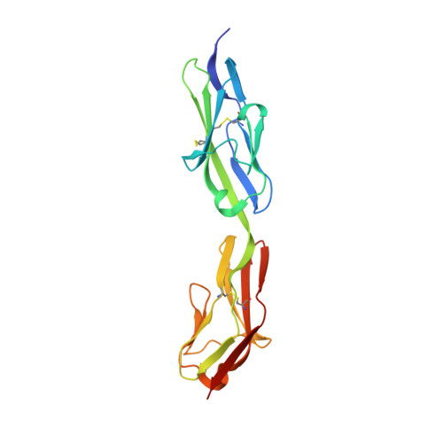
    Muscle-specific kinase (MuSK) is a receptor tyrosine kinase expressed exclusively in skeletal muscle, where it is required for formation of the neuromuscular junction. MuSK is activated by agrin, a neuron-derived heparan sulfate proteoglycan. Here, we report the crystal structure of the agrin-responsive first and second immunoglobulin-like domains (Ig1 and Ig2) of the MuSK ectodomain at 2.2 A resolution. The structure reveals that MuSK Ig1 and Ig2 are Ig-like domains of the I-set subfamily, which are configured in a linear, semi-rigid arrangement. In addition to the canonical internal disulfide bridge, Ig1 contains a second, solvent-exposed disulfide bridge, which our biochemical data indicate is critical for proper folding of Ig1 and processing of MuSK. Two Ig1-2 molecules form a non-crystallographic dimer that is mediated by a unique hydrophobic patch on the surface of Ig1. Biochemical analyses of MuSK mutants introduced into MuSK(-/-) myotubes demonstrate that residues in this hydrophobic patch are critical for agrin-induced MuSK activation.


  • Organizational Affiliation
    • Structural Biology Program, Skirball Institute of Biomolecular Medicine and Department of Pharmacology, New York University School of Medicine, New York, NY 10016, USA.

Macromolecule Content 

  • Total Structure Weight: 42.18 kDa 
  • Atom Count: 3,020 
  • Modeled Residue Count: 374 
  • Deposited Residue Count: 384 
  • Unique protein chains: 1

Macromolecules

Find similar proteins by:|  3D Structure
Entity ID: 1
MoleculeChains  Sequence LengthOrganismDetailsImage
Muscle-specific kinase receptor
A, B
192Rattus norvegicusMutation(s): 0 
Gene Names: Musk
EC: 2.7.10.1
UniProt
Find proteins for Q62838 (Rattus norvegicus)
Explore Q62838 
Go to UniProtKB:  Q62838
Entity Groups
Sequence Clusters30% Identity50% Identity70% Identity90% Identity95% Identity100% Identity
UniProt GroupQ62838
Sequence Annotations
Expand
Reference Sequence

Experimental Data & Validation

Experimental Data

  • Method: X-RAY DIFFRACTION
  • Resolution: 2.21 Å
  • R-Value Free:  0.258 (Depositor) 
  • R-Value Work:  0.216 (Depositor), 0.216 (DCC) 
  • R-Value Observed: 0.218 (Depositor) 
Space Group: P 21 21 2
Unit Cell:
Length ( Å )Angle ( ˚ )
a = 77.608α = 90
b = 118.004β = 90
c = 57.785γ = 90
Software Package:
Software NamePurpose
DENZOdata reduction
SCALEPACKdata scaling
REFMACrefinement
PDB_EXTRACTdata extraction
ADSCdata collection
HKL-2000data reduction
HKL-2000data scaling
AMoREphasing

Structure Validation

View Full Validation Report


Entry History 

Deposition Data

Revision History  (Full details and data files)

  • Version 1.0: 2006-11-28
    Type: Initial release
  • Version 1.1: 2007-10-16
    Changes: Version format compliance
  • Version 1.2: 2011-07-13
    Changes: Derived calculations, Version format compliance
  • Version 1.3: 2017-10-18
    Changes: Refinement description
  • Version 1.4: 2023-08-30
    Changes: Data collection, Database references, Derived calculations, Refinement description
  • Version 1.5: 2024-11-13
    Changes: Structure summary