2FX0

Crystal Structure of HlyIIR, a Hemolysin II transcriptional Regulator


Experimental Data Snapshot

  • Method: X-RAY DIFFRACTION
  • Resolution: 2.40 Å
  • R-Value Free: 0.276 
  • R-Value Work: 0.221 
  • R-Value Observed: 0.223 

wwPDB Validation   3D Report Full Report


This is version 1.2 of the entry. See complete history


Literature

Crystal Structure of Bacillus cereus HlyIIR, a Transcriptional Regulator of the Gene for Pore-forming Toxin Hemolysin II.

Kovalevskiy, O.V.Lebedev, A.A.Surin, A.K.Solonin, A.S.Antson, A.A.

(2007) J Mol Biol 365: 825-834

  • DOI: https://doi.org/10.1016/j.jmb.2006.10.074
  • Primary Citation of Related Structures:  
    2FX0

  • PubMed Abstract: 

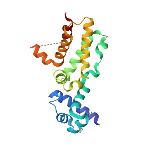
    Production of Bacillus cereus and Bacillus anthracis toxins is controlled by a number of transcriptional regulators. Here we report the crystal structure of B. cereus HlyIIR, a regulator of the gene encoding the pore-forming toxin hemolysin II. We show that HlyIIR forms a tight dimer with a fold and overall architecture similar to the TetR family of repressors. A remarkable feature of the structure is a large internal cavity with a volume of 550 A(3) suggesting that the activity of HlyIIR is modulated by binding of a ligand, which triggers the toxin production. Virtual ligand library screening shows that this pocket can accommodate compounds with molecular masses of up to 400-500 Da. Based on structural data and previous biochemical evidence, we propose a model for HlyIIR interaction with the DNA.


  • Organizational Affiliation

    Institute of Biochemistry and Physiology of Microorganisms, Russian Academy of Sciences, Pushchino, Moscow Region 142290, Russia.


Macromolecules
Find similar proteins by:  (by identity cutoff)  |  3D Structure
Entity ID: 1
MoleculeChains Sequence LengthOrganismDetailsImage
hemolysin II regulatory protein201Bacillus cereusMutation(s): 4 
Gene Names: hlyIIR
UniProt
Find proteins for Q7X506 (Bacillus cereus)
Explore Q7X506 
Go to UniProtKB:  Q7X506
Entity Groups  
Sequence Clusters30% Identity50% Identity70% Identity90% Identity95% Identity100% Identity
UniProt GroupQ7X506
Sequence Annotations
Expand
  • Reference Sequence
Small Molecules
Modified Residues  1 Unique
IDChains TypeFormula2D DiagramParent
MSE
Query on MSE
A
L-PEPTIDE LINKINGC5 H11 N O2 SeMET
Experimental Data & Validation

Experimental Data

  • Method: X-RAY DIFFRACTION
  • Resolution: 2.40 Å
  • R-Value Free: 0.276 
  • R-Value Work: 0.221 
  • R-Value Observed: 0.223 
  • Space Group: P 61 2 2
Unit Cell:
Length ( Å )Angle ( ˚ )
a = 124.208α = 90
b = 124.208β = 90
c = 79.667γ = 120
Software Package:
Software NamePurpose
REFMACrefinement
DENZOdata reduction
SCALEPACKdata scaling
SOLVEphasing

Structure Validation

View Full Validation Report


Entry History 

Deposition Data

Revision History  (Full details and data files)

  • Version 1.0: 2006-02-21
    Type: Initial release
  • Version 1.1: 2008-05-01
    Changes: Version format compliance
  • Version 1.2: 2011-07-13
    Changes: Advisory, Derived calculations, Refinement description, Version format compliance