2FNA | pdb_00002fna

Crystal structure of an archaeal aaa+ atpase (sso1545) from sulfolobus solfataricus p2 at 2.00 A resolution


Experimental Data Snapshot

  • Method: X-RAY DIFFRACTION
  • Resolution: 2.00 Å
  • R-Value Free: 
    0.226 (Depositor), 0.227 (DCC) 
  • R-Value Work: 
    0.174 (Depositor), 0.181 (DCC) 
  • R-Value Observed: 
    0.177 (Depositor) 

wwPDB Validation 3D Report Full Report

Validation slider image for 2FNA

Ligand Structure Quality Assessment 


This is version 1.4 of the entry. See complete history

Literature

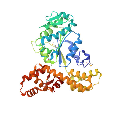
Crystal structure of a novel archaeal AAA+ ATPase SSO1545 from Sulfolobus solfataricus.

Xu, Q.Rife, C.L.Carlton, D.Miller, M.D.Krishna, S.S.Elsliger, M.A.Abdubek, P.Astakhova, T.Chiu, H.J.Clayton, T.Duan, L.Feuerhelm, J.Grzechnik, S.K.Hale, J.Han, G.W.Jaroszewski, L.Jin, K.K.Klock, H.E.Knuth, M.W.Kumar, A.McMullan, D.Morse, A.T.Nigoghossian, E.Okach, L.Oommachen, S.Paulsen, J.Reyes, R.van den Bedem, H.Hodgson, K.O.Wooley, J.Deacon, A.M.Godzik, A.Lesley, S.A.Wilson, I.A.

(2009) Proteins 74: 1041-1049

Macromolecule Content 

  • Total Structure Weight: 85.46 kDa 
  • Atom Count: 6,175 
  • Modeled Residue Count: 704 
  • Deposited Residue Count: 714 
  • Unique protein chains: 1

Macromolecules

Find similar proteins by:|  3D Structure
Entity ID: 1
MoleculeChains  Sequence LengthOrganismDetailsImage
Conserved hypothetical protein
A, B
357Saccharolobus solfataricus P2Mutation(s): 5 
Gene Names: 13814777
UniProt
Find proteins for Q97Y08 (Saccharolobus solfataricus (strain ATCC 35092 / DSM 1617 / JCM 11322 / P2))
Explore Q97Y08 
Go to UniProtKB:  Q97Y08
Entity Groups
Sequence Clusters30% Identity50% Identity70% Identity90% Identity95% Identity100% Identity
UniProt GroupQ97Y08
Sequence Annotations
Expand
Reference Sequence

Small Molecules

Ligands 3 Unique
IDChains Name / Formula / InChI Key2D Diagram3D Interactions
ADP

Query on ADP


Download:Ideal Coordinates CCD File
D [auth A],
L [auth B]
ADENOSINE-5'-DIPHOSPHATE
C10 H15 N5 O10 P2
XTWYTFMLZFPYCI-KQYNXXCUSA-N
EDO

Query on EDO


Download:Ideal Coordinates CCD File
E [auth A]
F [auth A]
G [auth A]
H [auth A]
I [auth A]
E [auth A],
F [auth A],
G [auth A],
H [auth A],
I [auth A],
J [auth A],
M [auth B]
1,2-ETHANEDIOL
C2 H6 O2
LYCAIKOWRPUZTN-UHFFFAOYSA-N
MG

Query on MG


Download:Ideal Coordinates CCD File
C [auth A],
K [auth B]
MAGNESIUM ION
Mg
JLVVSXFLKOJNIY-UHFFFAOYSA-N
Modified Residues  1 Unique
IDChains TypeFormula2D DiagramParent
MSE
Query on MSE
A, B
L-PEPTIDE LINKINGC5 H11 N O2 SeMET

Experimental Data & Validation

Experimental Data

  • Method: X-RAY DIFFRACTION
  • Resolution: 2.00 Å
  • R-Value Free:  0.226 (Depositor), 0.227 (DCC) 
  • R-Value Work:  0.174 (Depositor), 0.181 (DCC) 
  • R-Value Observed: 0.177 (Depositor) 
Space Group: P 1 21 1
Unit Cell:
Length ( Å )Angle ( ˚ )
a = 55.36α = 90
b = 108.35β = 100.31
c = 70.57γ = 90
Software Package:
Software NamePurpose
REFMACrefinement
XSCALEdata scaling
PDB_EXTRACTdata extraction
XDSdata reduction
SHELXphasing
SHARPphasing

Structure Validation

View Full Validation Report


Ligand Structure Quality Assessment 


Entry History 

Deposition Data

Revision History  (Full details and data files)

  • Version 1.0: 2006-02-07
    Type: Initial release
  • Version 1.1: 2008-05-01
    Changes: Version format compliance
  • Version 1.2: 2011-07-13
    Changes: Advisory, Source and taxonomy, Version format compliance
  • Version 1.3: 2023-01-25
    Changes: Database references, Derived calculations
  • Version 1.4: 2024-10-09
    Changes: Data collection, Structure summary