2BN2 | pdb_00002bn2

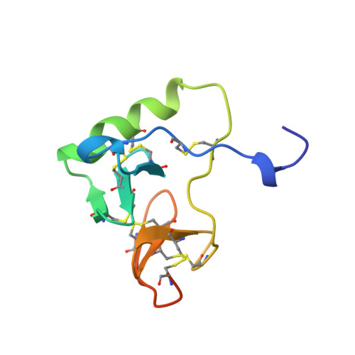
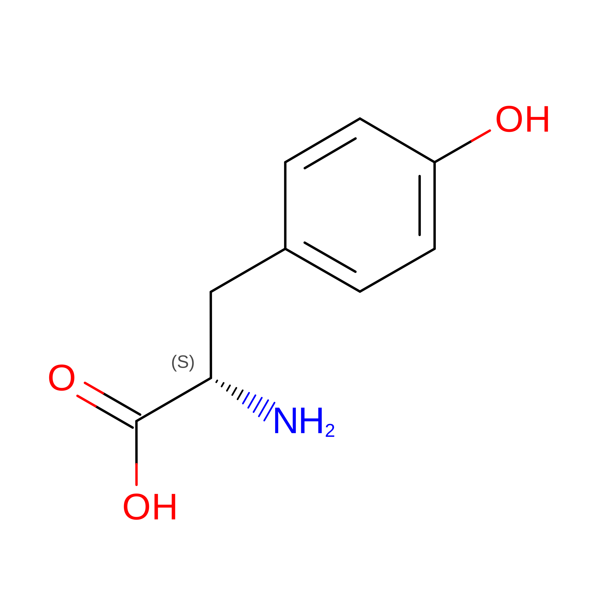
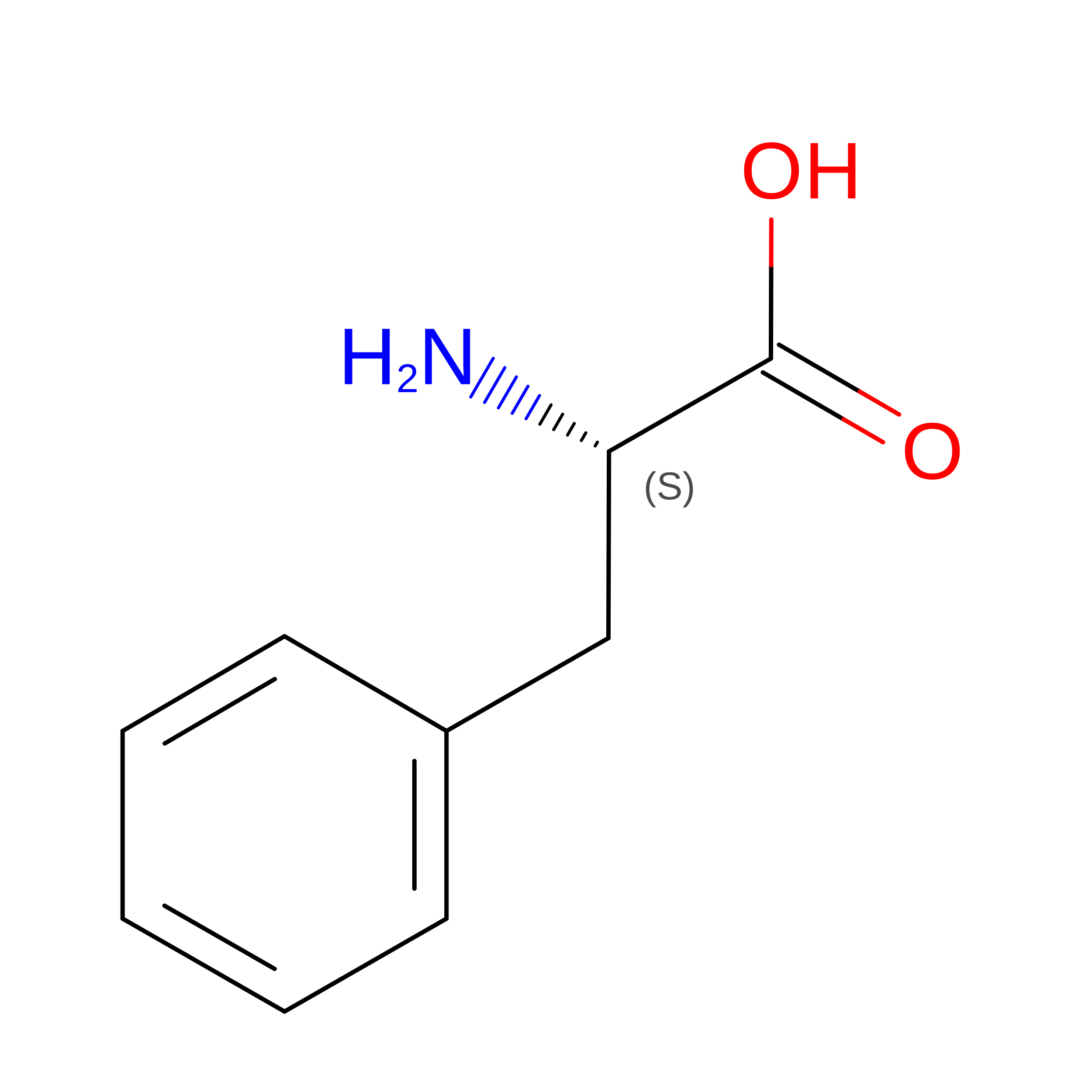
CRYSTAL STRUCTURE OF BOVINE NEUROPHYSIN II COMPLEXED WITH THE VASOPRESSIN ANALOGUE PHE-TYR AMIDE


Experimental Data Snapshot

  • Method: X-RAY DIFFRACTION
  • Resolution: 2.80 Å
  • R-Value Free: 
    0.262 (Depositor) 
  • R-Value Work: 
    0.208 (Depositor) 
  • R-Value Observed: 
    0.208 (Depositor) 

wwPDB Validation 3D Report Full Report

Validation slider image for 2BN2

This is version 1.4 of the entry. See complete history. 

Literature

Crystal structure of a bovine neurophysin II dipeptide complex at 2.8 A determined from the single-wavelength anomalous scattering signal of an incorporated iodine atom.

Chen, L.Q., Rose, J.P., Breslow, E., Yang, D., Chang, W.R., Furey Jr., W.F., Sax, M., Wang, B.C.

(1991) Proc Natl Acad Sci U S A 88: 4240-4244

  • DOI: https://doi.org/10.1073/pnas.88.10.4240
  • Primary Citation Related Structures: 
    2BN2

  • PubMed Abstract: 

    The crystal structure of a dipeptide complex of bovine neurophysin II has been solved at 2.8 A resolution solely by using single-wavelength anomalous scattering data from a single iodinated derivative. The asymmetric unit is an elongated tetramer of dimensions 110 x 40 x 30 A, composed of two dimers related by pseudo twofold symmetry. Each monomer consists of two homologous layers, each with four antiparallel beta-strands. The two regions are connected by a helix followed by a long loop. Monomer-monomer contacts involve antiparallel beta-sheet interactions, which form a dimer with two layers of eight beta-strands. One peptide per monomer occupies the principal hormone-binding pocket formed by part of the amino-terminal region and parts of the connecting helix and loop, with binding to protein consistent with conclusions drawn from solution studies. Dimer-dimer contacts involve the Tyr49 region adjacent to this site. A fifth dipeptide, of unknown biological significance, helps to stabilize one of the monomer-monomer interfaces and the tetramer-tetramer network in the crystal.


  • Organizational Affiliation: 
    • Department of Crystallography, University of Pittsburgh, PA 15260.

Macromolecule Content 

  • Total Structure Weight: 40.95 kDa 
  • Atom Count: 2,324 
  • Modeled Residue Count: 322 
  • Deposited Residue Count: 380 
  • Unique protein chains: 1

Macromolecules

Find similar proteins by:|  3D Structure
Entity ID: 1
MoleculeChains  Sequence LengthOrganismDetailsImage
NEUROPHYSIN IIA,
B [auth C],
C [auth E],
D [auth G]
95Bos taurusMutation(s): 0 
UniProt
Find proteins for P01180 (Bos taurus)
Explore P01180 
Go to UniProtKB:  P01180
Entity Groups
Sequence Clusters30% Identity50% Identity70% Identity90% Identity95% Identity100% Identity
UniProt GroupP01180
Sequence Annotations
Expand
Reference Sequence

Experimental Data & Validation

Experimental Data

  • Method: X-RAY DIFFRACTION
  • Resolution: 2.80 Å
  • R-Value Free:  0.262 (Depositor) 
  • R-Value Work:  0.208 (Depositor) 
  • R-Value Observed: 0.208 (Depositor) 
Space Group: P 21 21 21
Unit Cell:
Length ( Å )Angle ( ˚ )
a = 121.67α = 90
b = 67.96β = 90
c = 62.1γ = 90
Software Package:
Software NamePurpose
XENGENdata collection
XENGENdata reduction
ISASmodel building
X-PLORrefinement
XENGENdata scaling
ISASphasing

Structure Validation

View Full Validation Report


Entry History 

Deposition Data

Revision History  (Full details and data files)

  • Version 1.0: 1999-02-16
    Type: Initial release
  • Version 1.1: 2008-03-24
    Changes: Version format compliance
  • Version 1.2: 2011-07-13
    Changes: Version format compliance
  • Version 1.3: 2024-06-05
    Changes: Data collection, Database references, Derived calculations, Other
  • Version 1.4: 2024-10-30
    Changes: Structure summary